
Journal of Clinical Images and Medical Case Reports
ISSN 2766-7820
Case Report - Open Access, Volume 4
Contrast ultrasonography of nodular transformation of splenic sclerosing hemangioma: A case report
Fengyun Wu1; Tingting Lei1; Ting Sun1; Hualin Li2*
1Department of Medical Ultrasound, Qingdao Municipal Hospital (Group), Qingdao, Shandong 266000, China.
2Department of Medical Ultrasound, Zibo Maternal and Child Health Hospital, Zibo, Shandong 255029, China.
*Corresponding Author : Hualin Li
Department of Medical Ultrasound, Zibo Maternal
and Child Health Hospital, Zibo, Shandong 255029,
China.
Email: 287358887@qq.com
Received : Oct 17, 2023
Accepted : Nov 30, 2023
Published : Dec 07, 2023
Archived : www.jcimcr.org
Copyright : © Li H (2023).
Abstract
Citation: Wu F, Lei T, Sun T, Li H. Contrast ultrasonography of nodular transformation of splenic sclerosing hemangioma: A case report. J Clin Images Med Case Rep. 2023; 4(12): 2726.
Case data
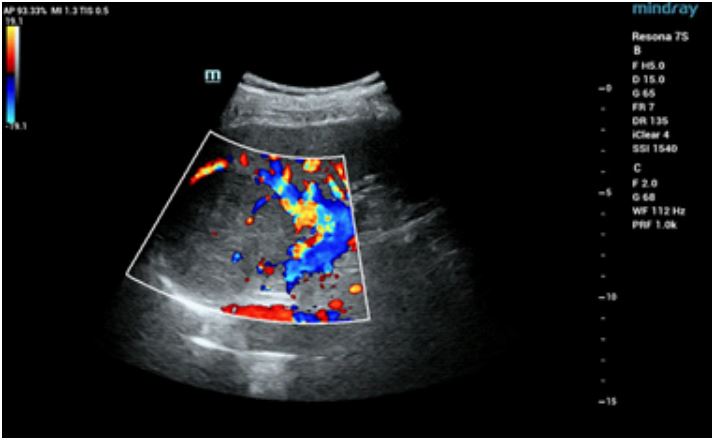
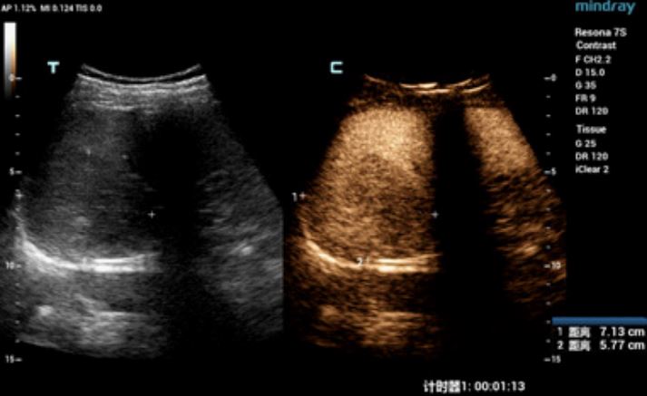
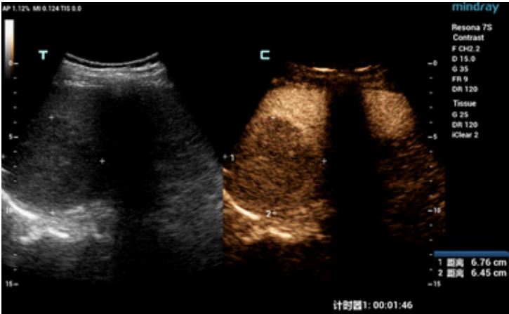
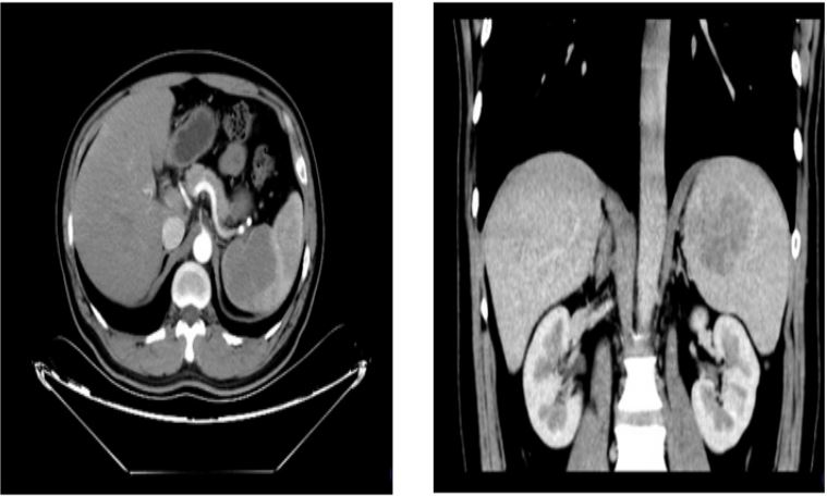
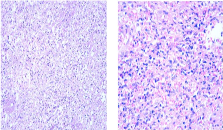
A 33-year-old male patient was admitted to our hospital due to splenomegaly and splenic space occupation on physical examination. Admission physical examination: general condition was normal, no fever, nausea, vomiting, abdominal distension, diarrhea and other symptoms, healthy in the past, no family history of disease. Ultra sonographic description: A single heterogeneous hypoechoic mass about 6.9×5.6 cm in size was detected in the lower pole of spleen. It was almost round in shape with clear boundary. A ring with slightly high echo was seen around the mass, and blood flow signals were seen in and around CDFI (Figure 1). Contrast Enhanced Ultrasound Sonography (CEUS) showed that the hypoechoic mass in the spleen presented concentric uneven and high enhancement in the arterial phase, with spokes in the center (Figure 2). In the venous phase, the contrast agent slowly withdrew, presenting low enhancement, with a size of about 7.6×6.5 cm after contrast, and the enhancement degree was always lower than that of the surrounding spleen parenchyma (Figure 3), Contrast-enhanced ultrasound suggested solid space-occupying lesions in the spleen, possibly benign lesions. In CT description, the spleen showed a rounded shadow with slightly low density, and the enhanced scan showed progressive enhancement, with a diameter of about 64 mm (Figure 4). CT prompt it as Lymphoma?. And malignant tumors are not excluded. Postoperative gross pathology showed: postoperative pathology revealed a nodular mass of 6x4.5x3 cm in size, with slightly hard gry-brown substance on the cut surface, partial scar area on the cut surface, with a little capsule, the mass was easily separated from the capsule and the surrounding splenic tissue, and pathological examination showed splenic sclerotic hemangiomatous nodular transformation (benign lesion) (Figure 5). CD31 immunohistochemical results: CD8 (+), (+), CD34 (+), Ki67 (+ 2%), CK (-), Vimentin (+), CD68 (+), SMA (+), CD21 (-), according to (-), CD1a (-). The patient’s prognosis is good.
Discussion
Transformation of the spleen sample nodular sclerosing hemangioma (sclerosing angiomatoid nodular transformations, SANT) originally called spleen capillary hemangioma or multiple nodules hemangioma, 2004 Marel etc. Put forward that the lesion is a benign vascular lesion. It was confirmed that it was named splenic sclerosing hemangiomatoid nodule transformation [1], which is a rare benign vascularized lesion unique to the spleen. It is often solitary, common in women, and can occur at any age, and is more common in 30-60 years old [2]. Due to its low incidence and non-specific clinical manifestations, it is often confused with diseases such as hemangioma, hamartoma and lymphoma. At present, there are no large sample studies reported, and most cases are reported. There are no reports about the CEUS manifestations of this disease, and the CEUS features of this case are summarized as follows.
Conventional ultrasound of SANT mostly shows equal or low echo, and the internal echo is mostly uninform, and there is high echo in different numbers in the shape of cords or small pieces, which is caused by the thick fibrous sclerosis tissue enclosed and separated by hemangiomatous nodules in the lesion, and there are few cystic bleeding or calcification [3]. In this case, conventional ultrasound showed a low-echo mass with circular and clear boundary, uneven echo, and visible blood flow signals in and around CDFI. CEUS showed high concentric uneven enhancement in the arterial phase, with hub-shaped enhancement in the center, and low enhancement in the venous phase with slow withdrawal of contrast agent, which was always lower than the surrounding spleen parenchyma. CT enhancement of this disease is mainly manifested by early enhancement of the edge and interior of the lesion in the arterial phase, and continuous cardioid enhancement in the portal vein phase and delayed phase, most of which are accompanied by obvious “spokes sign”. No obvious necrotic cystic changes are observed in the lesion, and the boundary becomes clear after enhancement [4]. Common enhancement modes in CT include progressive and centripetal enhancement, spoke-like or nodular enhancement, and scarring of central delayed enhancement fibers [5]. The pattern of enhancement in this case is consistent with progressive enhancement with central spoke enhancement
The pathological characteristics of SANT are the formation of multiple hemangiomatoid nodules of different sizes mainly distributed in the fibrous sclerosis stroma, and arranged in the inner “spokes” [4]. Its hemangiomatoid nodules are composed of splenic red pulp components with clear microscopic boundaries [6]. The cause is still under investigation. SANT tends to be a benign hyperplastic lesion rather than a tumor, and is a reactive vascular lesion that can be cured by surgery [7]. Correctly identifying the nature of spleen space occupying disease is crucial. The contrast-enhanced ultrasound features of this case can provide some basis for diagnosis. However, as SANT is a sporadic tumor, its pathogenesis and prognosis need to be further studied.
Foundation: 2020 Medical Research Guiding Plan of Qingdao city 2020-WJZD018
References
- Martel M, Cheuk W, Lombardi L, et al. Sclerosing angiomatoid nodular transformation (SANT): Report of 25 cases of a distinctive benign splenic lesion [J]. The American journal of surgical pathology, 2004 28: 1268-1279.
- Vigorito R, Scaramuzza D, Pellegrinelli A, et al. Sclerosing angiomatoid nodular transformation (SANT) of the spleen: A case report on CT and MRI [J]. BJR Case Rep. 2019; 5: 20180036.
- Xiujuan Zhang, Zhikui Chen, Qingfu QIAN, Huipin Chen, Linyan Tong, et al. Ultrasonographic findings of nodular transformation of spleen sclerosing hemangioma. Chinese journal of medical imaging technology. 2019; 35: 1053-1056.
- Feng Zhao, Zhoupeng Ma. CT diagnosis and differentiation of splenic sclerosing hemangiomatous nodular transformation. Journal of hepatobiliary and pancreatic surgery. 2020; 32: 484-487.
- Barat M, Hoeffel C, Aissaoui M, et al. Focal splenic lesions: Imaging spectrum of diseases on CT, MRI and PET/CT [J]. Diagnostic and Interventional Imaging. 2021; 389-396.
- Silver DS, Pointer DT Jr, Slakey DP. Solid Tumors of the Spleen: Evaluation and Management. J Am Coll Surg. 2017; 224: 1104-1111.
- Rommel Ojeda, Gabriel A Molina, Galo E Jiménez, Hernán González, Johanna C Pinto, et al. Sclerosing Angiomatoid Nodular Transformation (SANT) of the spleen: A rare cause of acute abdomen, Journal of Surgical Case Reports. 2021; 126: 112-117.