
Journal of Clinical Images and Medical Case Reports
ISSN 2766-7820
Clinical Image - Open Access, Volume 4
Multimodal imaging evaluation of femoral exostosis in a pediatric patient: A radiation-free approach
Marco Quadrelli1*; Tommaso Baccaglini1; Aldo Morra2
1Synlab Euganea Medica Diagnostic Center, Italy.
2IRCCS SDN Synlab S.p.A., Italy.
*Corresponding Author : Marco Quadrelli
Synlab Euganea Medica Diagnostic Center, Italy.
Email: dr.marcoquadrelli@gmail.com
Received : Nov 22, 2023
Accepted : Dec 19, 2023
Published : Dec 26, 2023
Archived : www.jcimcr.org
Copyright : © Quadrelli M (2023).
Citation: Quadrelli M, Baccaglini T, Morra A. Multimodal imaging evaluation of femoral exostosis in a pediatric patient: A radiation-free approach. J Clin Images Med Case Rep. 2023; 4(12): 2756.
Description
A 12-year-old girl presented with left knee pain following an accidental fall. Physical examination revealed local pain and restricted joint movement. The patient had no significant prior medical conditions or traumas.
Differential diagnosis ruled out musculoskeletal injuries and other orthopedic conditions.
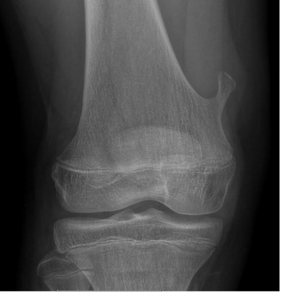
Bilateral comparative knee radiography identified an exostosis at the distal third of the right femur incidentally.
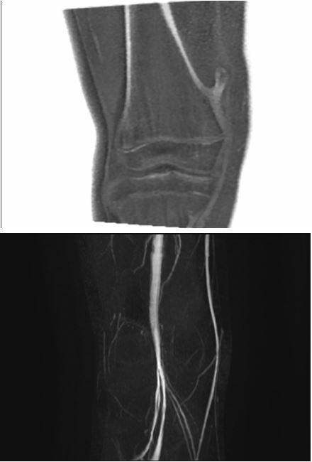
To assess the relationship between the exostosis and blood vessels, lower limb CT angiography with contrast was performed. To explore the possibility of conducting future followups without exposing the patient to ionizing radiation, experimental MRI sequences oZTEo and TOF without contrast were employed to evaluate the exostosis and adjacent structures without ionizing radiation exposure.
This multimodal imaging approach facilitated a comprehensive assessment of the exostosis and surrounding tissues using less invasive imaging techniques.
The bilateral comparative knee radiograph incidentally discovered an exostosis at the distal third of the right femur. This image vividly displays the bony prominence, offering insight into its location and appearance.
Utilizing MRI sequences oZTEo and TOF without contrast provided a detailed view of the exostosis and adjacent structures. These images illuminate the lesion’s characteristics and its proximity to blood vessels, presenting a non-ionizing radiation approach for comprehensive analysis.
These images exemplify a multimodal diagnostic approach, enabling a thorough assessment of the femoral exostosis in a pediatric patient while avoiding ionizing radiation exposure.
References
- Murphey MD, Choi JJ, Kransdorf MJ, Flemming DJ, Gannon FH. Imaging of osteochondroma: variants and complications with radiologic-pathologic correlation. Radiographics. 2000; 20(5): 1407-1434. [PubMed] (https://pubs.rsna.org/doi/full/10.1148/ radiographics.20.5.g00se081407)
- Ramos R, Cugat R, Cuscó X, Seijas R, Ares O, García-Balletbó M. Osteochondromas about the knee in the pediatric and adolescent population. J Pediatr Orthop. 2010; 30(2): 188-192. [PubMed](https://pubmed.ncbi.nlm.nih.gov/20179567/)
- Rozanes I, Ciampi P, Borrello A, Rossi F, Cappellani F, Indovina A. Magnetic resonance imaging of osteochondromas and exostoses: A pictorial review. Acta Biomed. 2018; 89(4- S): 30-39. [PubMed](https://pubmed.ncbi.nlm.nih.gov/30561458/)