
Journal of Clinical Images and Medical Case Reports
ISSN 2766-7820
Clinical Image - Open Access, Volume 5
A rare case of extra-parotid pleomorphic adenoma
Hann-Ziong Yueh; Shih-Chun Lu; Tzu-Ying Chen*
Department of Otolaryngology, Taipei Medical University Hospital, Taipei, Taiwan.
*Corresponding Author : Tsu-Ying Chen
Department of Otolaryngology, Taipei Medical University Hospital, Taipei, Taiwan.
Email: tziyingchen@gmail.com
Received : Feb 23, 2024
Accepted : Mar 07, 2024
Published : Mar 14, 2024
Archived : www.jcimcr.org
Copyright : © Tzu-Ying C (2024).
Citation: Hann-Ziong Y, Shih-Chun L, Tzu-Ying C. A rare case of extra-parotid pleomorphic adenoma. J Clin Images Med Case Rep. 2024; 5(3): 2922.
Description
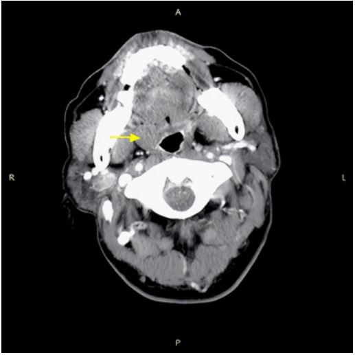
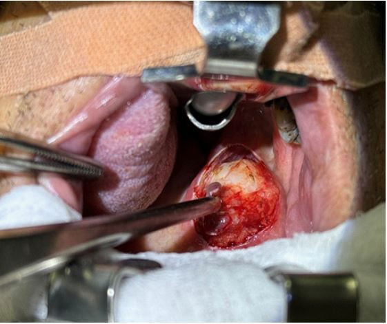
A 56-year-old male patient presented to the Department of Otolaryngology with a gradually enlarging soft palatal mass on the right side over several months. Clinical examination revealed a non-ulcerated, dome-shaped, palatal swelling on the soft palate. The mass, measuring approximately 2x2 cm, was smooth, uninodular, firm, non-tender, and non-fluctuant, without involvement of adjacent teeth. No overlying ulceration or discoloration was observed. Computed tomography scan indicated a well-circumscribed solid lesion (2.3 x 1.9 x 1.7 cm) with no evidence of infiltration into surrounding soft-tissue structures, the pterygopalatine fossa, or palatine nerve foramina (Figure 1, arrow). The lesion was successfully resected through a transoral approach, easily dissected from surrounding tissue, and sparing the overlying mucosa (Figure 2). Pathological analysis confirmed a diagnosis of pleomorphic adenoma. The patient recovered well, and subsequent follow-up in the outpatient department revealed no signs of recurrent tumor. Pleomorphic adenoma is the most common neoplasm of the large salivary glands and happens to be the commonest one constituting about 70% of which 84% occur in parotid, 8% in the submandibular gland and 4-6% in the minor salivary glands. Corresponding to small glands, a small minority of tumors are also located in the palate, lip, oral/nasal cavity and neck region [1,2].
References
- Pires FR, Pringle GA, de Almeida OP, Chen SY. Intra-oral minor salivary gland tumors: a clinicopathological study of 546 cases. Oral Oncol. May 2007; 43(5): 463-70. doi: 10.1016/j.oraloncology.2006.04.008
- Pinkston JA, Cole P. Incidence rates of salivary gland tumors: results from a population-based study. Otolaryngol Head Neck Surg. Jun 1999; 120(6): 834-40. doi:10.1016/S0194- 5998(99)70323-2.