Journal of Clinical Images and Medical Case Reports
ISSN 2766-7820
Clinical Image - Open Access, Volume 5
Symptomatic vallecular cyst
Hassan Doulhousne1,3*; Ilias Benchafai2,3; Badr Slioui1,3; Salah Belasri1,3; El Mehdi Atmane1,3; Abdelilah Mouhsine1,3;Nabil Hammoune1,3
1Department of Radiology, Avicenne Military University Hospital, Marrakech, Morocco.
2Department of Ear, Nose and Throat, Avicenne Military University Hospital, Marrakech, Morocco.
3Faculty of Medicine and Pharmacy, Cadi Ayyad University, Marrakech, Morocco.
*Corresponding Author : Hassan Doulhousne
Department of Radiology, Avicenne Military University Hospital, Marrakech, Morocco.
Email: ha.doulhousne@uca.ac.ma
Received : Feb 23, 2024
Accepted : Mar 08, 2024
Published : Mar 15, 2024
Archived : www.jcimcr.org
Copyright : © Doulhousne H (2024).
Citation: Doulhousne H, Benchafa I, Slioui B, Belasri S, Atmane EM, et al. Symptomatic vallecular cyst. J Clin Images Med Case Rep. 2024; 5(3): 2924.
Description
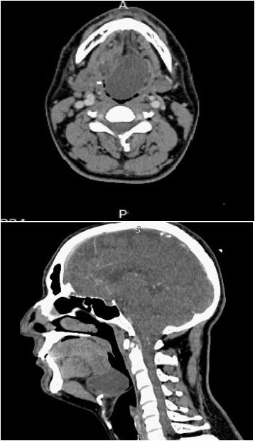
We report the case of a 32-year-old man suffering from a progressive dysphagia 4 months ago, who was seen in an ENT consultation and whose endoscopic examination showed a large shiny formation at the base of the tongue, considerably reducing the pharyngeal lumen. Cervical computed tomography (CT) showed a homogeneous cystic formation filling the vallecula and pushing down the epiglottis. The patient was diagnosed with vallecular cyst (Figure 1). The treatment was endoscopic, with marsupialization of the cyst after aspiration of its contents.