Journal of Clinical Images and Medical Case Reports
ISSN 2766-7820
Clinical Image - Open Access, Volume 5
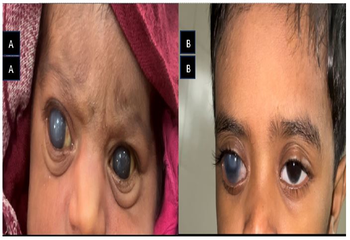
Capturing a child with congenital glaucoma
Kiran GC1*; Diwash Sunar2
1Lecturer, Department of Ophthalmology, Nobel Medical College Teaching Hospital, Biratnagar, Nepal.
2Lecturer, Department of ENT, Birat Medical College Teaching Hospital, Biratnagar, Nepal.
*Corresponding Author : Kiran GC
Lecturer, Department of Ophthalmology, Nobel Medical College Teaching Hospital, Biratnagar, Nepal.
Email: gc.kiran31@gmail.com
Received : Mar 29, 2024
Accepted : Apr 23, 2024
Published : Apr 30, 2024
Archived : www.jcimcr.org
Copyright : © Kiran GC (2024).
Citation: Kiran GC, Sunar D. Capturing a child with congenital glaucoma. J Clin Images Med Case Rep. 2024; 5(4): 3019.
Description
Congenital glaucoma, though rare, is a sight threatening condition which can cause lifelong impact to a child’s life. It often presents at birth, arises from abnormal development of the eye’s drainage system, leading to increased pressure within the eye. Without timely intervention, it can result in irreversible vision loss.
Parents may observe excessive tearing, sensitivity to light, and enlarged cloudy corneas which are the telltale signs of Glaucoma. These manifestations are often mistaken for common eye irritations, so it is also necessary for clinicians to recognise signs of Glaucoma for early referral. Delayed diagnosis risks irreversible vision loss. Treatment typically involves medical and surgical intervention to improve drainage and alleviate pressure.