Journal of Clinical Images and Medical Case Reports
ISSN 2766-7820
Clinical Image - Open Access, Volume 5
Unveiling the hidden: Point-of-Care Ultrasound (POCUS) reveals the diagnosis of rectus sheath hematoma
*Corresponding Author : Sundas Sajid
Aga Khan University, Pakistan.
Email: sundus.sajid@aku.edu
Received : May 23, 2024
Accepted : Jun 04, 2024
Published : Jun 11, 2024
Archived : www.jcimcr.org
Copyright : © Sajid S (2024).
Citation: Sajid S. Unveiling the hidden: Point-of-Care Ultrasound (POCUS) reveals the diagnosis of rectus sheath hematoma. J Clin Images Med Case Rep. 2024; 5(6): 3108.
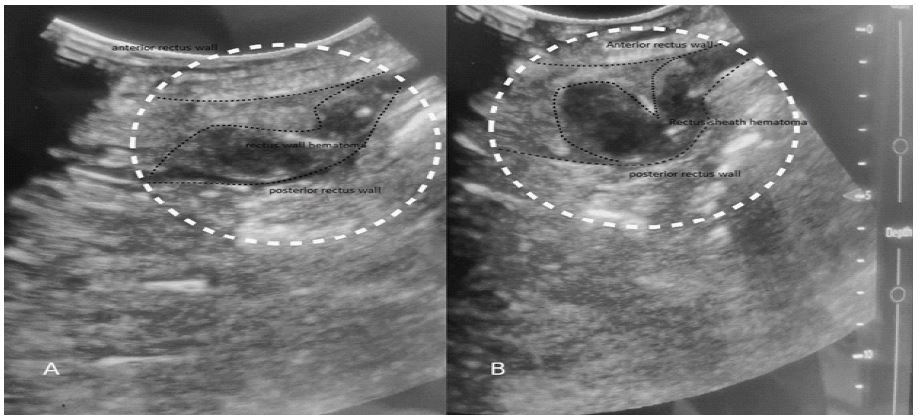
Description
A 42-year-old female with abdominal pain and on anticoagulation presented with an acute Rectus Sheath Hematoma (RSH), a rare abdominal pathology. Point-of-Care Ultrasonography (POCUS) was performed at the bedside, revealing a hypoechoic lesion in the left rectus sheath causing mass effect, consistent with RSH. The ultrasound image depicts the hematoma as a dark area within the rectus sheath, demonstrating the utility of POCUS in rapid and noninvasive diagnosis. Conservative management was pursued, leading to excellent outcomes. This case underscores the importance of POCUS as a first-line imaging tool for diagnosing RSH, enabling real-time assessment of hematoma size, extent, and progression. Knowledge of sonographic features is crucial for timely diagnosis and preventing life-threatening complications. POCUS facilitates expedited conservative management of RSH, optimizing patient outcomes.