Journal of Clinical Images and Medical Case Reports
ISSN 2766-7820
Clinical Image - Open Access, Volume 5
Unfolding an intriguing theory of an 18 week old malformed foetus
Alushika Jain1*; Roohi Gupta2
1Junior Resident, Radiodiagnosis Department, Datta Meghe Institute of Education and Research, Wardha, India.
2Assitant Professor, Radiodiagnosis Department, Datta Meghe Institute of Education and Research, Wardha, India.
*Corresponding Author : Alushika Jain
Junior Resident, MD Radiodiagnosis Department, Datta Meghe Institute of Education and Research, Wardha, India.
Email: alushika23@gmail.com
Received : May 20, 2024
Accepted : Jun 05, 2024
Published : Jun 12, 2024
Archived : www.jcimcr.org
Copyright : © Jain A (2024).
Citation: Jain A, Gupta R. Unfolding an intriguing theory of an 18 week old malformed foetus. J Clin Images Med Case Rep. 2024; 5(6): 3114.
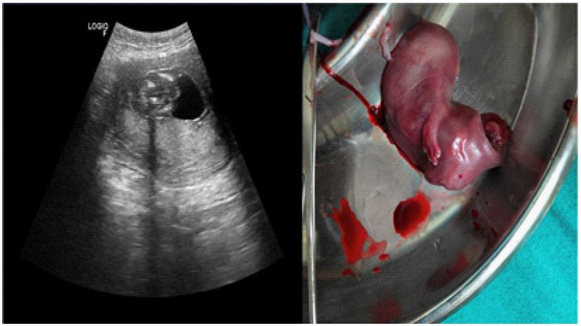
Description
An 18-week pregnant female walked into our antenatal care for her first scan. On doing the first scan, it was evident that the foetus was malformed because the fetal limbs were untraceable, and also, the whole foetus was crumpled like a ball. The sac and the foetus were small and inconsistent with an 18-week pregnancy. Although there was cardiac activity in the foetus, the anatomical structures were not formed properly. The ultrasound revealed a large, translucent mass near the fetal neck region, resembling a fluid-filled sac measuring approx 3.8 x 1.8 cm with altered curvature of the spine. The lower limbs and upper limbs were not appreciated. The patient also had severe oligohydramnios with a single deepest pocket measuring 1.4 cm. The placenta was posterior and reached up to the internal os. A provisional diagnosis of cystic hygroma or meningocele was given. Surrounding tissues appear compressed or displaced by the expanding cystic hygroma. The image depicts a distinct anomaly indicative of cystic hygroma, requiring further evaluation and management. Firstly, the location of the meningocele is crucial. Situated at the occipital region, it appears as a sac-like protrusion through the fetal skull. This sac contains meninges, the protective membranes covering the brain and spinal cord, and potentially cerebrospinal fluid. Its appearance may vary from translucent to opaque, depending on its contents. The image depicts an aborted 18-week foetus having cystic hygroma and no outer skull, also resembling a seal with no lower limbs and short upper limbs. This not only points out how important it is to get regular antenatal scans but also helps in the early detection and diagnosis of various anomalies for appropriate management and counselling of affected families at a very early period. Hence, counselling the patient in antenatal care is very important regarding ultrasonography.