
Journal of Clinical Images and Medical Case Reports
ISSN 2766-7820
Clinical Image - Open Access, Volume 5
A clinical image of rare presentation of advanced diabetic wound
*Corresponding Author : Dipika Chakole
Assistant Professor, Department of Kayachikitsa, MGACHRC, Datta Meghe Institute of Higher Education and Research, Sawangi (M), Wardha, Maharashtra, India.
Email: dchakole16@gmail.com
Received : May 22, 2024
Accepted : Jun 05, 2024
Published : Jun 12, 2024
Archived : www.jcimcr.org
Copyright : © Chakole D (2024).
Keywords: Diabetic foot; Gangrened toes; T2DM.
Citation: Chakole D. A clinical image of rare presentation of advanced diabetic wound. J Clin Images Med Case Rep. 2024; 5(6): 3115.
Introduction and objective
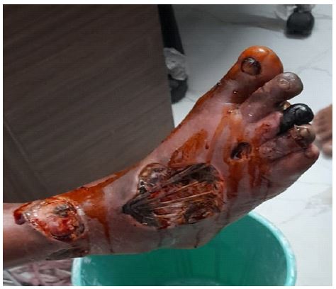
A diabetes mellitus is one of the most commonly occurred non communicable disease which affects approximately 422 million peoples worldwide. The mortality of 2 million people per year is estimated due to diabetes and its complication [1]. Diabetic foot is most commonly found chronic complication of Diabetes and leading indication of prolonged hospital stay for its management. Diabetic foot has classical tried of Neuropathy, ischemia and infection and became very complicated scenario to treat. In the pathophysiological event of diabetic foot, soft tissue infection caused due conditions like neuropathy and trauma with peripheral artery occlusive disease [2]. This clinical image is presentation of a 65 years old male patient visited to Clinic with Bandaged foot. Having complaints of Wound over foot (in the last 3-4 months), fever with chills (since 20-25 days). A patient was diagnosed with T2DM since 10-15 years and prescribed with oral anti diabetic Medicine. Due to lack of awareness and poor financial condition patient didn’t take medicine properly and didn’t monitor blood sugar level regularly. When Patient was suffered with simple wound at right foot, he didn’t visit any physician for prolonged time and results into this presentation. Patient was immediately shifted to higher facility centre for further management.
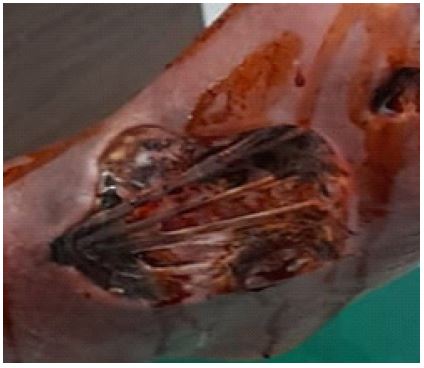
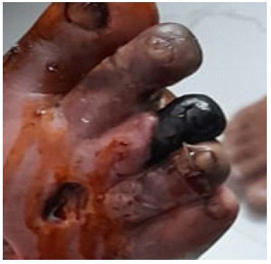
Image descriptions: Figure 1 is a clinical image picture of presentation of wound in the patient. In this image a clear presentation exposed extensor digitorum longus muscle is seen. 2nd and 3rd toes both are gangrened; a round wound patch is also present at ankle region. Generally diabetic foot condition occurred at palmer region but in this it presented in dorsal region with advanced condition exposing internal muscle tendons and gangrened toes.
References
- GBD 2019 Diabetes in the Americas Collaborators. Burden of diabetes and hyperglycaemia in adults in the Americas, 1990-2019: a systematic analysis for the Global Burden of Disease Study 2019. Lancet Diabetes Endocrinol. 2022; 10(9): 655-667. doi:10.1016/S2213-8587(22)00186-3.
- Bandyk DF. The diabetic foot: Pathophysiology, evaluation, and treatment. Semin Vasc Surg. 2018; 31(2-4): 43-48. doi: 10.1053/j.semvascsurg.2019.02.001.