
Journal of Clinical Images and Medical Case Reports
ISSN 2766-7820
Clinical Image - Open Access, Volume 5
Ocular coloboma with microphtalmia
Taibi Ouiam*; Bouanane R; Sqalli A; Belkouchi L; N Allali N; El Haddad S; Chat L
Department of Radiology, National Institute of Oncology, Rabat, Morocco.
*Corresponding Author : Taibi Ouiam
Department of Radiology, National Institute of Oncology, Rabat, Morocco.
Email: ouiam.tb@gmail.com
Received : Jun 04, 2024
Accepted : Jun 19, 2024
Published : Jun 26, 2024
Archived : www.jcimcr.org
Copyright : © Ouiam T (2024).
Citation: Ouiam T, Bouanane R, Sqalli A, Belkouchi L, Allali NN, et al. Ocular coloboma with microphtalmia. J Clin Images Med Case Rep. 2024; 5(6): 3140.
Clinical image
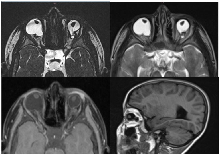
A 3-year-old child was referred for small eyes and always keeping them closed. MRI revealed dysmorphic ocular globes with loss of their spherical shape and a cystic polilobulated lesion communicating with the globe; hypointense on T1-weighted images and hyperintense on T2-weighted images without contrast enhancement associated with a microphthalmia. Findings are suggestive of a retrobulbar colobomatous congenital cyst (Figure 1).
Discussion
A coloboma is a developmental abnormality resulting from the failure of closure of the embryonic choroidal fissure, that occurs between gestational days 35 and 41. Presenting as a cleft that may involve the retina, choroid, sclera, iris, ciliary body, lens, or optic nerve head. A coloboma can be an isolated finding in an otherwise healthy individual or part of a complex malformation syndrome of known or unknown etiology [1]. Colobomas may be associated with reduced ocular globe size or with normal size and are bilateral in more than 60% of cases [2]. Both sexes are equally affected [3].
On MRI, these cysts vary in size and appear as hypointense on T1-weighted images and hyperintense on T2-weighted images. The exact communication site between the cyst and the globe may not always be clear on imaging and the differential diagnosis to consider in imaging is staphyloma [4].
References
- Gujar SK, Gandhi D. Congenital Malformations of the Orbit. Neuroimaging Clin N Am. 2011; 21(3): 585-602.
- Pagon RA Ocular coloboma. Surv Ophthalmol. 1981 25: 223-236.
- Kindler P Morning glory syndrome: unusual congenital optic disc anomaly. Am J Ophthalmo. 1970; 169: 376-384.
- Pahwa S, Sharma S, Das CJ, Dhamija E, Agrawal S. Intra-orbital Cystic Lesions: An Imaging Spectrum. Curr Probl Diagn Radiol. 2015; 44(5): 437-448.