
Journal of Clinical Images and Medical Case Reports
ISSN 2766-7820
Clinical Image - Open Access, Volume 5
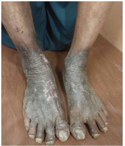
Severe case of atopic dermatitis: An image
Ashwin Gokhare1*; Kartikesh Gokhare2
1Department of Panchakarma, Mahatma Gandhi Ayurved College Hospital and Research Centre, Datta Meghe Institute of Higher Education & Research, Sawangi, Wardha, Maharashtra, India.
2General Physician, Diploma in Homeopathic Medicine and Surgery, Certificate Course in Modern Pharmacology (CCMP), Chandrapur, Maharashtra, India.
*Corresponding Author : Ashwin Gokhare, MD
Department of Panchakarma, Mahatma Gandhi Ayurved College Hospital and Research Centre, Datta Meghe Institute of Higher Education & Research, Sawangi, Wardha, Maharashtra, India.
Email: gokhareashwin@gmail.com
Received : Jun 05, 2024
Accepted : Jun 21, 2024
Published : Jun 28, 2024
Archived : www.jcimcr.org
Copyright : © Gokhare A (2024).
Keywords: Eczema; Contact dermatitis; Dermatology; Skin disease.
Citation: Gokhare A, Gokhare K. Severe case of atopic dermatitis: An image. J Clin Images Med Case Rep. 2024; 5(6): 3144.
Introduction
Atopic dermatitis, a particular type of eczema, stands as the prevalent chronic inflammatory skin condition [1]. Patients with atopic dermatitis possess a flawed skin barrier, making them prone to dryness and susceptible to environmental triggers and allergens. This vulnerability leads to inflammation, itching, and the characteristic clinical signs of atopic dermatitis. The barrier deficiency may arise partly from reduced levels of ceramides, essential lipids in the outer skin layer that uphold the barrier’s function and prevent water loss. This compromised barrier allows allergens and irritants to enter, triggering an exaggerated immune response-Th2 in acute cases (with increased IL-4, and IL-5 cytokines) and Th1 in chronic instances (with IFN-gamma and IL-12). Scratching aggravates the situation by prompting skin cells to release inflammatory molecules like TNF-alpha, IL-1, and IL-6. Additionally, decreased levels of antimicrobial peptides in the skin contribute to the colonization of Staphylococcus aureus, observed in over 90% of atopic dermatitis patients. S. aureus colonization can exacerbate inflammation, leading to secondary infections and impetigo in atopic dermatitis lesions [2].
The research found that in certain nations, more than 20% of children experience AD, yet its occurrence widely differs across the globe. Among 6-7-year-olds, AD prevalence spans from 0.9% in India to 22.5% in Ecuador. Recent findings indicate elevated rates in Asia and Latin America.
Case report
A male patient, age 45 with complaints of severe itching and watery discharge at bilateral ankle and foot region & darkening of skin at same region came to OPD. The patient was a farmer by occupation and used to work in water-logged soil for almost 2-3 hours daily [3].
Progression of disease: The patient states that 4-5 years ago he started getting small pustules over the ankle region with watery discharge. Over time he states that itching gradually increased. Slowly started seeing the darkening of the skin over the region. After ignoring the disease, he came to OPD 1 year ago.
Diagnosis: Atopic dermatitis.
Differential diagnosis: Lichen simplex, Lichen Planus, Psoriasis, Tinea.
Treatment: We started with plain plain antibiotic Tab. Azithromycin 500 OD for 5 days. Antihistamine Tab. Bilastin 20 mg OD HS for 15 days. Tab. Deflazacort 6 mg 1 tab BD for 5 days. For local application, Betamethasone Valerate and neomycin cream were given.
Follow-up after 15 days symptoms like itching were reduced, and after 30 days he got relief from watery discharge, and itching with drying of the dermatitis.
References
- Paller A, Jaworski JC, Simpson EL, Boguniewicz M, Russell JJ, Block JK, Tofte S, Dunn JD, Feldman SR, Clark AR, Schwartz G, Eichenfield LF. Major Comorbidities of Atopic Dermatitis: Beyond Allergic Disorders. Am J Clin Dermatol. 2018; 19(6): 821-838.
- Kolb L, Ferrer-Bruker SJ. Atopic Dermatitis. [Updated 2023 Aug 8]. In: StatPearls [Internet]. Treasure Island (FL): StatPearls Publishing; 2023. Available from: https://www.ncbi.nlm.nih.gov/books/NBK448071/.
- Sophie Nutten; Atopic Dermatitis: Global Epidemiology and Risk Factors. 2015; 66(1): 8-16. https://doi.org/10.1159/000370220.