
Journal of Clinical Images and Medical Case Reports
ISSN 2766-7820
Case Report - Open Access, Volume 5
The puzzle of idiopathic clubbing and alcoholic pancreatitis: A case report
Parmendra Sirohi1; Manaswi Vishwakarma2*; Rahul Gupta3; Peeyush Sharma3; Abhishek Verma3
1MD (General Medicine), Senior Professor, Sardar Patel Medical College and PBM Hospitals, Bikaner, India.
2MBBS, Sardar Patel Medical College and PBM Hospitals, Bikaner, India.
3PG Resident (General Medicine), Sardar Patel Medical College and PBM Hospitals, Bikaner, India.
*Corresponding Author : Manaswi Vishwakarma
Sardar Patel Medical College and PBM Hospitals, Pawanpuri, Bikaner, India.
Tel: +91-9414334356;
Email: manaswivishwakarma@gmail.com
Received : May 28, 2024
Accepted : Jul 05, 2024
Published : Jul 12, 2024
Archived : www.jcimcr.org
Copyright : © Vishwakarma M (2024).
Abstract
Clubbing of the digits has been recognized as a clinical sign of variety of diseases from earliest of the times. Digital clubbing was first ever reported in a patients with empyema by Hippocrates himself. Since then, digital clubbing has been associated with various underlying pulmonary, cardiovascular, neoplastic, infectious, hepatobiliary, endocrine, and gastrointestinal diseases. Primary or idiopathic clubbing is very rare presentation in which the features of clubbing are seen even in the absence of any recognizable associated disease. And surprisingly, the specific underlying patho-physiological mechanism associated with primary and even secondary clubbing remains in the shadows till date. Many theories have been proposed, yet none of them claimed the acknowledgement and acceptance as an comprehensive explanation for the phenomenon. The motives of this article extend beyond the clinical case, influencing and promoting future research to comprehensively study the phenomenon of clubbing and the not-so-known mechanisms behind it; further helping us to formulate differentials, diagnostics and prevention strategies.
Keywords: Acute pancreatitis; Aesthetic medicine; Idiopathic; Lung cancer; Nail clubbing.
Citation: Sirohi P, Vishwakarma M, Gupta R, Sharma P, Verma A. The puzzle of idiopathic clubbing and alcoholic pancreatitis: A case report. J Clin Images Med Case Rep. 2024; 5(7): 3166.
Introduction
Clubbing of the nails is soft tissue swelling of the terminal phalanx, resulting in flattening the angle between the nail bed and the nail. The conditions related with digital clubbing are pulmonary causes (75-80%), cardiovascular causes (10-15%), gastrointestinal and hepatobiliary causes (5-15%) and miscellaneous causes (5-15%) [1]. On rare occasions, digital clubbing may be primary or idiopathic, which seen in the absence of any apparent known condition [2]. There are numerous etiologies which results in clubbing and all the patients with an underlying disease do not necessarily show clubbing. Thus estimating the incidence of digital clubbing pose challenges. Roughly 1% of all internal medicine admissions shows sign of clubbing and 40% of and connective tissue changes. High-resolution magnetic resonance imaging using a contrast agent was done in a study by Nakamura in patients digital clubbing and healthy volunteers. Nail bed hyper-vascularization was seen with clubbed nails [5,6].
Case report
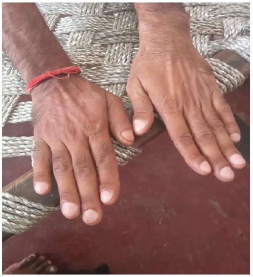
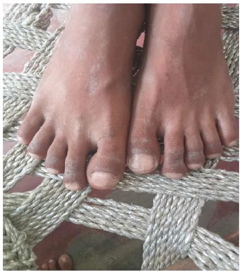
A 41 year old male presented to us with complaint of pain abdomen since last 8 days, localized to epigastric region, penetrating in nature, radiating to back, increased with food intake and relieved with medication [7]. It was associated with nausea without vomiting. There is no history of loose stools, steatorrhea, hematemesis or malena. Patient had history of alcohol ingestion few hours before development of symptoms. No history of similar episode in the past with no history of any systemic illness. Patient gave vague history of bulbous enlargement of fingers first noticed by his mother 8 years back but didn’t seek any medical attention for it; and not present during birth and early childhood. There was no history of cyanotic spells, recurrent chest infections, chronic cough or hemoptysis, jaundice, bloody diarrhea or altered bowel habits. Our patient was chronic alcoholic since last 5 years taking around 30-40 gram of alcohol per day. On examination, patient was alert, conscious and well oriented to time, place and person. Blood pressure was 110/70 mm of Hg in right upper limb, pulse rate of 84 bpm, and oxygen saturation of 97% in room air. There was no evidence of pallor, icterus, cyanosis or lymphadenopathy. Examination of hand and feet revealed bulbous enlargement of distal phalanxes identified as Grade 3 clubbing (Figurs 1 & 2). No localized tenderness, skin thickening or joint swelling was found. On GI examination patient had epigastric tenderness without palpable liver or spleen. Respiratory examination revealed decrease air entry with crackles in left lower zone. Rest of the system examination were within normal limits.
Comprehensive blood count revealed leukocytosis with counts of 17330 which was predominantly neutrophilic, mild anemia with Hb of 11.6 g/dl with macrocytosis (MCV 104 fl). Liver function and renal function tests were normal. Thyroid function test was normal.
Serum amylase level were 384 IU/l and lipase were 786 IU/l, corroborating our suspicion of acute pancreatitis (alcohol induced).
USG whole abdomen showed bulky pancreatic tail with peripancreatic & perinephric fat inflammation with normal liver size and echo-texture. Contrast CT abdomen was performed after 48 hrs which revealed moderate acute pancreatitis with tail end necrosis and adjacent fluid collection which confirmed our diagnosis of acute necrotic pancreatitis however no clues were found regarding GI causes of clubbing. HRCT thorax revealed mild left pleural effusion (secondary to pancreatitis) without any evidence of fibrosis, bronchiectatic changes, mass or lung abscess. To rule out cardiac causes of clubbing, trans thoracic echo-cardiography was performed which did not reveal any clots, vegetation or any structural heart defect. Patient was kept nil per mouth, treated with i.v. fluids and antibiotics; as his abdominal symptoms alleviated oral feeding was initiated. Our patient was counseled for regular follow up visits in order to identify any abnormal disease process that may manifest as time passes and to treat it all the same.
Discussion
Digital clubbing is examined during initial evaluation of the patient. There are various possible etiologies of clubbing, thus a thorough history with an intent to recognize the characteristic features and cause of clubbing should be taken into account. The history is followed by the physical exam, where nail clubbing is assessed. Acquired clubbing is most often associated with pulmonary or cardiovascular diseases, so initial workup especially targeting these systems is a very reasonable approach. Furthermore, to make a diagnosis of idiopathic clubbing rigorous in-depth examination, work ups, and investigations must be done to eliminate any known possible cause of secondary clubbing.
Differential diagnosis of nail clubbing includes: Neoplastic diseases like lung cancer, mesothelioma, lymphoma, nasopharyngeal carcinoma, and esophageal cancer. Other non-neoplastic heart and lung related conditions like aortic aneurysm, bronchiectasis, lung abscess, cyanotic congenital heart diseases, cystic fibrosis, sarcoidosis, empyema, pulmonary fibrosis, endocarditis. Other conditions like celiac disease, ascariasis, Inflammatory bowel disease, thyroid disorders are also related. Many attempts have been made by number of researchers to determine the underlying mechanism of clubbing [8]. On critically analyzing their attempts, increased blood flow to distal part of digits resulting from vasodilation comes out to be a common factor in development of clubbing. But currently it is not agreed upon whether this vasodilation is due to local or circulating vasodilators, or due to hypoxia, or due to some neural (especially vagal) component, or a combination of these and other mediators. We could not establish any significant history of this being present by birth in the patient or any recurrent pulmonary infection. None of the parents nor the siblings had clubbing. Systemic examination of respiratory and cardiovascular system ruled out majority of the causes which was further consolidated by CT and Echocardiography findings.
Since our patient noted this bulbous enlargement just few years back, not present in childhood we believe it to be acquired clubbing which could not be ascertained to any known cause during our investigation, henceforth “idiopathic” in nature. Furthermore, the history and the investigations eliminates the possibility of alcohol induced pancreatitis to be the cause of clubbing. Alcohol induced pancreatitis is not a known cause of clubbing, therefore it prompts us to indulge in more comprehensive research related to clubbing and its causes.
Aesthetic changes due to digital clubbing in an individual can lead to complications especially surrounding the psycho-social aspects of day-to-day life. There has been several reports indicating that acquired clubbing is reversible with targeted intervention of the underlying cause, although the data is very scarce [9]. Thus identification of the root cause of clubbing remains of utmost importance, as the prognosis and intervention needed depends upon the it.
Conclusion
In conclusion, digital clubbing is a physical exam finding which often correlates with a serious underlying disease. Clubbing can be recognized by medical students, nurses, and physicians conducting the physical examination. Regardless of who identifies the clubbing first, an inter-professional team approach is the ideal way to evaluate and manage a patient with clubbing. Furthermore, cases of clubbing are warehouse of unknown information and have a great potential for further comprehensive researches, therefore must not remain unrecognized.
References
- Burcovschii S, Aboeed A. Nail Clubbing. [Updated 2022 Sep 24]. In: StatPearls [Internet]. Treasure Island (FL): StatPearls Publishing. 2024. Available from: https://www.ncbi.nlm.nih.gov/books/NBK539713/#.
- Friedman HH. Clubbing. In: Friedman, M.H., Ed., Problem-Oriented Medical Diagnosis, 7th Edition, Lipponcpott Williams and Wilkins, Philadelphia. 2001; 277-278.
- Vandemergel X, Renneboog B. Prevalence, aetiologies and significance of clubbing in a department of general internal medicine. Eur J Intern Med. 2008; 19(5): 325-9. doi: 10.1016/j.ejim.2007.05.015. Epub 2008 Feb 11. PMID: 18549933.
- Dickinson CJ. The aetiology of clubbing and hypertrophic osteoarthropathy. Eur J Clin Invest. 1993; 23(6): 330-8. doi:10.1111/j.1365-2362.1993.tb02032.x. PMID: 8344332.
- Tucker JR. Nail Deformities and Injuries. Prim Care. 2015; 42(4): 677-91. doi: 10.1016/j.pop.2015.08.005. PMID: 26612379.
- Baran R. Sémiologie unguéale [The use of nails to diagnosis diseases]. Presse Med. 2014; 43(11): 1208-15. French. doi: 10.1016/j.lpm.2014.06.014. PMID: 25443634.
- Nakamura J, Halliday NA, Fukuba E, Radjenovic A, Tanner SF, Emery P, et al. The Microanatomic Basis of Finger Clubbing -- A High-resolution Magnetic Resonance Imaging Study. J Rheumatol. 2014; 41(3): 523-7.
- Burcovschii S, Aboeed A. Nail Clubbing. [Updated 2022 Sep 24]. In: StatPearls [Internet]. Treasure Island (FL): StatPearls Publishing. 2024. Available from: https://www.ncbi.nlm.nih.gov/books/NBK539713/#.
- McPhee SJ. Clubbing. In: Walker HK, Hall WD, Hurst JW, editors. Clinical Methods: The History, Physical, and Laboratory Examinations. 3rd ed. Boston: Butterworths; 1990; 44. PMID: 21250207.