Journal of Clinical Images and Medical Case Reports
ISSN 2766-7820
Clinical Image - Open Access, Volume 5
Multi-modality cardiac imaging in an adult patient with unrepaired tetralogy of fallot
*Corresponding Author : Jed Valentiner Shrewsbury
Faculty of Medicine, Imperial College London, London,
United Kingdom.
Tel: 07739527333; Email: jed.shrewsbury1@nhs.net
Received : Oct 10, 2024
Accepted : Oct 30, 2024
Published : Nov 06, 2024
Archived : www.jcimcr.org
Copyright : © Valentiner Shrewsbury J (2024).
Citation: Valentiner Shrewsbury J. Multi-modality cardiac imaging in an adult patient with unrepaired tetralogy of fallot. J Clin Images Med Case Rep. 2024; 5(11): 3329.
Description
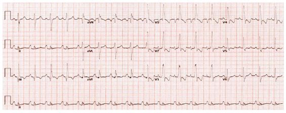
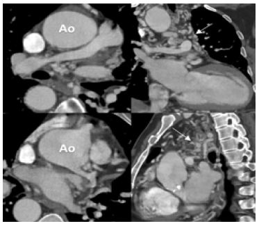
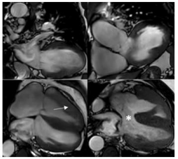
Tetralogy of Fallot (TOF) is the most common cyanotic congenital heart defect in children worldwide. Surgical correction is required in early childhood and non-operative survival past the fourth decade is rarely described. We report an interesting case demonstrating the utility of multimodality imaging, including Transthoracic Echocardiography (TTE), Computed Tomography Pulmonary Angiography (CTPA) and Cardiac Magnetic Resonance imaging (CMR), in the assessment of a symptomatic patient in his 40s with unrepaired TOF. Together, these three imaging modalities provided complementary information of the patient’s cardiac and valvular anatomy, morphology and function. CTPA was performed to accurately visualise the confluence of the pulmonary arteries which TTE and CMR had failed to do, revealing extensive systemic-pulmonary artery collateral vessels and pulmonary atresia. These investigations enabled more accurate prognostication and facilitated further planning of management.