Journal of Clinical Images and Medical Case Reports
ISSN 2766-7820
Clinical Image - Open Access, Volume 5
The fortuitous discovery of bilateral retinal coloboma
Latifa Sbai*; Romaissae Benkirane; Taha Boutaj; Zineb Hilali; Hamza Lazaar; Salma Hamidi; Samira Tachfouti; Lalla Ouafa Cherkaoui
Ophthamology Department A, Ibn Sina University Hospital Center, Mohamed V Souissi University, Rabat, Morocco.
*Corresponding Author : Latifa Sbai
Ophthamology Department A, Ibn Sina University Hospital Center, Mohamed V Souissi University, Rabat, Morocco.
Email: latifasbai95@gmail.com
Received : Nov 03, 2024
Accepted : Nov 21, 2024
Published : Nov 28, 2024
Archived : www.jcimcr.org
Copyright : © Sbai L (2024).
Keywords: Coloboma; Retina; Papilla; OCT; Fluorescein Angiography.
Citation: Sbai L, Benkirane R, Boutaj T, Hilali Z, Lazaar H, et al. The fortuitous discovery of bilateral retinal coloboma. J Clin Images Med Case Rep. 2024; 5(11): 3361.
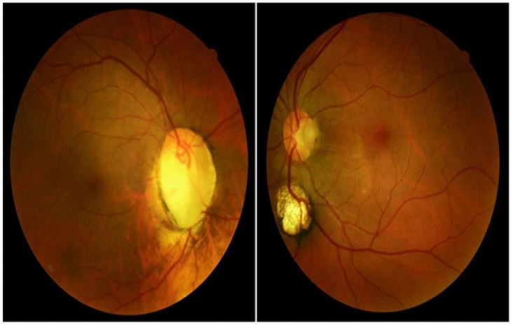
Description
Colobomas are congenital malformations due to an abnormality in the closure of the colobomic cleft during embryonic life, which can affect various ocular structures such as the retina. We report the case of a 50-year-old female patient with no previous history of the condition, who presented for a change of optical correction. Uncorrected visual acuity was 8/10 ODG, with normal ocular tone. Examination of the anterior segment was unremarkable. The dilated fundus revealed a papillary coloboma with a papillary diameter on the right, and a sub-papillary chorioretinal coloboma with a papillary diameter on the left. The rest of the retinal examination did not reveal any associated abnormality (Figure 1). Papillary OCT revealed significant loss of fibres and RNFL on the right, with a mean thickness of 53 μm. As part of the malformative assessment, an ECG, renal ultrasound and brain imaging were requested, all of which came back normal.