
Journal of Clinical Images and Medical Case Reports
ISSN 2766-7820
Clinical Image - Open Access, Volume 5
A rare case of primary synovial chondromatosis of unilateral knee joint in female: A clinical image
*Corresponding Author : Manisha kapadiya
Assistant Professor, Department of Shalyatantra, JS Ayurved Mahavidhyalaya, Nadiad- 387001, Gujarat, India.
Tel: +91-9974047713;
Email: manisha.kapadia15@gmail.com
Received : Nov 01, 2024
Accepted : Nov 22, 2024
Published : Nov 29, 2024
Archived : www.jcimcr.org
Copyright : © Kapadiya M (2024).
Citation: Kapadiya M. A rare case of primary synovial chondromatosis of unilateral knee joint in female: A clinical image. J Clin Images Med Case Rep. 2024; 5(11): 3366
Description
Synovial Chondromatosis (SC) is a rare begin condition that can rarely be converted into malignancy. This condition is related to the synovial membrane of joints, bursa, and tendon sheath. It can be primary or secondary. Primary synovial chondromatosis is of unknown etiology. Removal of loose bodies provides symptomatic relief and there is a chance of recurrence. So synovectomy with the removal of loose bodies is treatment of choice [1].
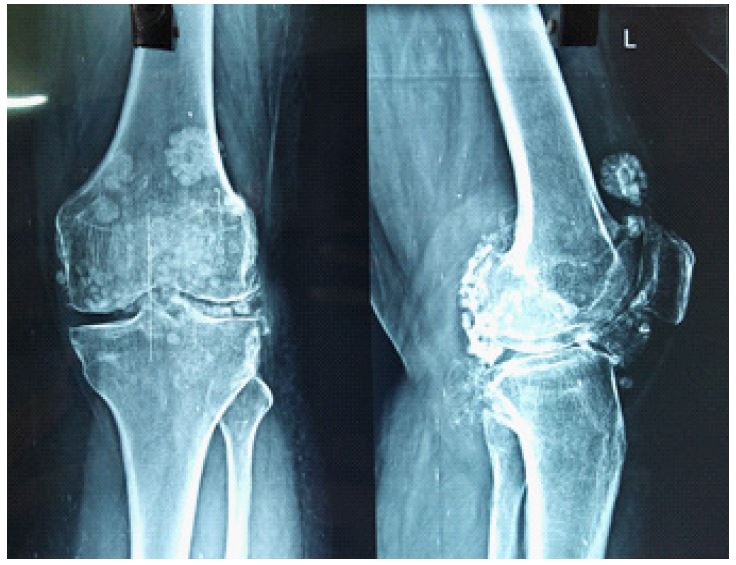
A 37 year old non diabetic non hypertensive female, housewife presented with left knee joint pain and swelling for the last 3 years. There was a restricted range of motion of the left knee joint. There was no history of trauma or injury to the left knee joint. There was no history of fever or discharge from the joint. On examination, diffuse swelling involving the left knee, non-erythematous, soft, non-tender, with crepitus and palpable hard nodules of varying size. His knee flexion was limited to 50°. X-ray of the affected left knee joint revealed multiple intra-articular loose radio-opaque chondroid bodies of different sizes and shapes with preserved tibia-femoral space. Visualized bones appreciated degenerative changes in the form of endplate sclerosis, patella spiking, and osteophyte growth. This case diagnosed as primary synovial chondromatosis on the basis of radiological findings as well as clinical findings.
References
- Chaliyadan S, Gujar A, Vallikkad S, Kataria R. Synovial Chondromatosis in a Rural Healthcare Setting. Cureus. 2023; 5(2): e34498. Doi: 10.7759/cureus.34498.