
Journal of Clinical Images and Medical Case Reports
ISSN 2766-7820
Clinical Image - Open Access, Volume 6
Clinical image: Supraorbital venous pulsation due to severe tricuspid regurgitation
Cooper B Kersey, MD*; Margaret L Isaac, MD
Department of Medicine, Division of Cardiology, University of Washington Seattle, USA.
*Corresponding Author : Cooper B Kersey
Department of Medicine, Division of Cardiology,
University of Washington Seattle, 1959 Pacific St
Seattle, WA 98195, USA.
Email: kerseycb@uw.edu
Received : Jan 13, 2025
Accepted : Feb 12, 2025
Published : Feb 19, 2025
Archived : www.jcimcr.org
Copyright : © Kersey CB (2025).
Citation: Kersey CB, Isaac ML. Clinical image: Supraorbital venous pulsation due to severe tricuspid regurgitation. J Clin Images Med Case Rep. 2025; 6(2): 3474.
Description
An 88-year-old man with a history of nonischemic cardiomyopathy and atrial fibrillation presented after an acute syncopal episode in the setting of subacute dyspnea and weight gain. On exam, a grade 2/6 holosystolic murmur at the left sternal border and prominent jugular veins were noted. Closer examination of his internal jugular vein revealed that the venous pulsations were most prominent in systole with a prominent CV wave. He had evidence of venous pulsations to the level of his supraorbital vein at 30o in bed and the pattern of pulsations was irregularly irregular, consistent with his known atrial fibrillation. A transthoracic echocardiogram revealed severe, centrally directed tricuspid regurgitation due to failure of coaptation of the tricuspid valve leaflets secondary to right ventricular dilatation.
The bedside examination of jugular venous pressure is challenging and studies have demonstrated disagreement between direct measurements of central venous pressure and clinician estimates [1]. The assessment of the jugular venous pulse is made more difficult when the patient has tricuspid valve pathology or atrial fibrillation. The physiologic jugular venous pulse is made up of three positive waveforms which are named “a”, “c”, and “v” waves. The “a” wave results from atrial contraction into the right ventricle and is thus absent in patients who are in atrial fibrillation. The “c” wave is caused by isovolumetric contraction of the right ventricle against the tricuspid valve and the “v” wave represents the passive filling of the right atrium. In tricuspid regurgitation, as seen with our patient, the “c” and “v” waves merge to form a prominent “CV” wave which is a singular broad waveform [2].
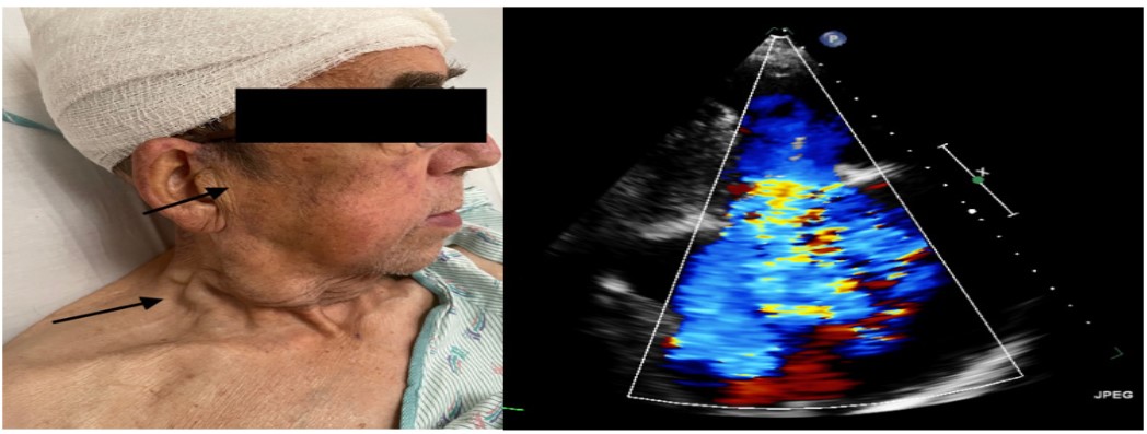
Video 1 (supplementary file): Jugular venous and supraorbital venous distension with prominent CV wave due to severe tricuspid regurgitation. Note the irregular irregularity of the venous pulsations due to the underlying cardiac rhythm of atrial fibrillation.
An 88-year-old man with a history of nonischemic cardiomyopathy and atrial fibrillation presented after an acute syncopal episode in the setting of subacute dyspnea and weight gain. On exam a grade 2/6 holosystolic murmur at the left sternal border and prominent jugular veins were noted. Closer examination of his internal jugular vein revealed that the venous pulsations were most prominent in systole (C wave) and coupled with a prominent V wave. He had evidence of venous pulsation to the level of his supraorbital vein at 30o in bed and the pattern of pulsations was irregularly irregular, consistent with his known atrial fibrillation. The transthoracic echocardiogram ordered for his syncope work-up revealed severe, centrally directed tricuspid regurgitation due to failure of coaptation of the tricuspid valve leaflets secondary to the severity of his right ventricular dilatation. He is being considered for outpatient percutaneous tricuspid valve repair.
References
- McGee SR. Physical examination of venous pressure: a critical review. Am Heart J. 1998; 136: 10-8.
- Applefeld MM. The Jugular Venous Pressure and Pulse Contour. In: Walker HK, Hall WD, Hurst JW, editors. Clinical Methods: The History, Physical, and Laboratory Examinations. 3rd edition. Boston: Butterworths. 1990.