
Journal of Clinical Images and Medical Case Reports
ISSN 2766-7820
Clinical Image - Open Access, Volume 6
Tendon xanthomata in a 68-year-old woman with primary biliary cholangitis-related hyperlipidemia
Savvoula Savvidou, MD, MSc, PhD*
Department of Internal Medicine, Hepatology Outpatients Clinic, “G. Gennimatas” General Hospital of Thessaloniki, Greece.
*Corresponding Author : Savvoula Savvidou
Department of Internal Medicine, Hepatology
Outpatients Clinic, “G. Gennimatas” General
Hospital of Thessaloniki, 41 Ethnikis Aminis Street,
ZIP code 54635, Thessaloniki, Greece.
Tel: +306977774301;
Email: ssavidou22@gmail.com
Received : Jan 27, 2025
Accepted : Feb 17, 2025
Published : Feb 24, 2025
Archived : www.jcimcr.org
Copyright : © Savvidou S (2025).
Abstract
Skin xanthomata form firm-to-hard, subcutaneous nodules, with normal overlying skin, that usually grow slowly around tendons, consisting of foamy cells derived from lipid-laden macrophages. Even though tendon xanthomata are highly prevalent in familial syndromes of hypercholesterolemia, secondary causes may result in their formation. In this case, we present a 68-year-old woman with extensive tendon xanthomata in her hands, induced by primary biliary cholangitis - related lipoprotein-X accumulation.
Keywords: xanthoma; hyperlipidemia; hypercholesterolemia; lipoprotein X; primary biliary cholangitis; primary biliary cirrhosis; cholestasis.
Abbreviations: PBC: primary biliary cholangitis; UDCA: ursodeoxycholic acid; t.i.d.: ter in die (three times a day); q.d.: quaquedia (once a day); AMA: anti-mitochondrial autoantibodies; ALP: alkaline phosphatase; γ-GT: γ-glutamyl-transpeptidase; HDL: high-density lipoprotein; LDL: low-density lipoprotein; Ig: immunoglobulin.
Citation: Savvidou S. Tendon xanthomata in a 68-year-old woman with primary biliary cholangitis-related hyperlipidemia. J Clin Images Med Case Rep. 2025; 6(2): 3481.
Case presentation
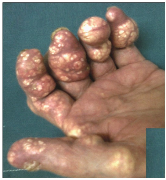
A 68-year-old woman with a history of primary biliary cholangitis (PBC) presented to the Hepatology Outpatients Clinic for routine follow-up. She had been diagnosed with PBC six years ago, and was under ursodeoxycholic acid (UDCA) 250 mg t.i.d., atorvastatin/ezetimibe 20/10 mg q.d., and vitamin D3 25.000 IU per week. Diagnosis of PBC, as said, had been based on abnormal hepatic biochemistry and positive anti-mitochondrial autoantibodies (AMA), but no laboratory reports were available at the time of the visit. The patient reported never having any symptoms of fatigue or itch. On clinical examination, a painless enlarged liver was detected, with no other signs of chronic liver disease (absence of jaundice, ascites, splenomegaly). Interestingly, the patient had extensive tendon xanthomata on both of her hands (Figure 1), dated since her middle age and stable in size for the past 10 years. No xanthelasmas were noted. Family history was unrevealing. Routine laboratory tests were requested, as well as an upper right quadrant ultrasonography with shear wave elastography. Alkaline Phosphatase (ALP) was 220 mg/dL (normal <120), γ-glutamyl-transpeptidase (γGT) 98 mg/dL (normal <55), total bilirubin 2,2 mg/dL (normal <1), aminotransferases and platelet count within normal, and severe hyperlipidemia despite standard hypolipidemic treatment; total cholesterol was 382 mg/dL, triglycerides 125 mg/dL, high-density lipoprotein (HDL) 32 mg/dL, and low-density lipopro- tein (LDL) 325 mg/dL. AMA was positive in a titer of 1:40, with measurements of serum immunoglobulins (IgM, IgG) within normal. All other additional testing was negative. Ultrasonography showed a bright liver, uncomplicated cholelithiasis, and a normal-sized spleen. Liver stiffness was measured on an average of 8.6 kPa (±1.8). Patient was advised to increase UDCA dosage to 1.250 mg per day, in order to comply with the PBC recommended dose of 13-15 mg/kg/day [1], while atorvastatin/ezetimib and vitamin D3 continued as they were. A bone density scan was requested for her next visit in three months.
Discussion
Skin xanthomata are yellowish (xanthos = yellow/blond in Greek), firm-to-hard, subcutaneous nodules, with normal overlying skin, that usually grow slowly around tendons [2]. They were first described histologically in the early 1900s as benign lesions, consisting of foamy cells derived from lipid-laden macrophages [2,3]. Even though the exact formation mechanism is still unclear, presence of xanthomata is beyond doubt indicative of metabolic lipid disturbance –the so-called dislipidosis - that involves not only cholesterol, but other fractions as well, such as triglycerides, phospholipids or un-esterified fatty acids [2,3]. Both xanthelasmas and xanthomata are highly prevalent in cases of severe hyperlipidemia like familial homozygous hypercholesterolemia. Nevertheless, secondary causes of hyperlipidemia like cholestasis-related conditions may lead to xanthoma formation [4]. In this setting, PBC is a chronic autoimmune cholestatic liver disease, associated with hypercholesterolemia in 75-95% of cases [5]. However, xanthomata in patients with cholestasis (although rare, estimated in less than 5% of cases) usually arise from excessive accumulation of lipoprotein X that lacks atherogenic properties [4,5]. Patients with PBC and hypercholesterolemia should be evaluated individually for their risk for cardiovascular disease [1,5], and statin use is therefore recommended, although not solely sufficient to normalize lipid levels.
Declarations
Ethics: Written informed consent for publication was obtained from the patient.
Generative AI: No AI-assisted technologies were used for this publication.
Conflict of interest: No competing financial interests related to this report.
References
- Hirschfield GM, Dyson JK, Alexander GJM, Chapman MH, Collier J, Hübscher S, et al. The British Society of Gastroenterology/ UK-PBC primary biliary cholangitis treatment and management guidelines. Gut. 2018; 67: 1568-94.
- Nezafati KA, Cruz P. Xanthoma, tendon. Dermatology advisor, https://www.dermatologyadvisor.com/home/decision-supportin-medicine/dermatology/xanthoma-tendon/. 2019.
- Fleischmajer R. Cutaneous and tendon xanthomas. Dermatologica. 1964; 128: 113-32.
- Ashorobi D, Liao H. Lipoprotein X-Induced Hyperlipidemia. In: StatPearls. Treasure Island (FL): StatPearls Publishing. 2024. https://pubmed.ncbi.nlm.nih.gov/34283509.
- Wah-Suarez MI, Danford CJ, Patwardhan VR, Jiang ZG, Bonder A. Hyperlipidaemia in primary biliary cholangitis: treatment, safety and efficacy. Frontline Gastroenterol. 2019; 10: 401-8.