
Journal of Clinical Images and Medical Case Reports
ISSN 2766-7820
Clinical Image - Open Access, Volume 6
Adenoma malignum: A rare uterine malignancy
*Corresponding Author : Charanjeet Singh
Assistant Professor, Yale University School of
Medicine, USA.
Email: charanjeet.singh@yale.edu
Received : Jan 22, 2025
Accepted : Feb 26, 2025
Published : Mar 05, 2025
Archived : www.jcimcr.org
Copyright : © Charanjeet S (2025).
Citation: Charnjeet S. Adenoma malignum: A rare uterine malignancy. J Clin Images Med Case Rep. 2025; 6(3): 3496.
Description
A 71-year-old G2P2 female with a history of HIV, hypertension, pre diabetes, obesity, and sleep apnea presents with multiple episodes of vaginal bleeding, abdominal pain and unintentional weight loos for two months.
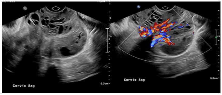
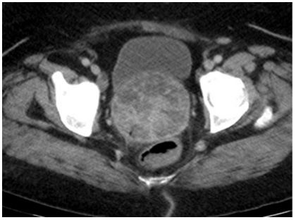
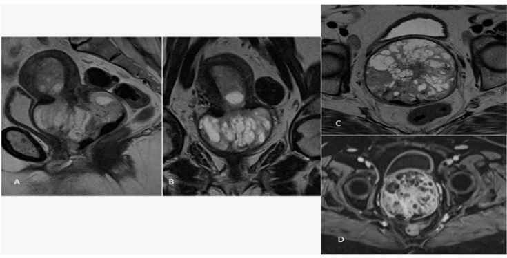
A transvaginal and transpelvic ultrasound was performed, which demonstrated a lobulated heterogenous multicystic-solid mass with internal vascularity in the region of cervix. Based on the imaging findings imaging diagnosis of Adenoma Malignum was suggested. She underwent CT scan and MRI pelvis confirming the findings of a lobulated heterogenous multicystic-solid mass in the lower uterine segment/cervix with extension into the adjacent vaginal wall. There was suggestion of parametrial invasion and enlarged lymph nodes along pelvic walls and at aortic bifurcation.
She underwent neoadjuvant chemotherapy followed by hysterectomy, bilateral salpingo-oophorectomy, distal omentectomy, lymph node dissections, posterior vaginal resection and tumor debulking.
The histopathology report confirmed this to be Adenoma Malignum, also known as minimal deviation adenocarcinoma, is a rare subtype of mucinous adenocarcinoma of the cervix [1- 4]. Its prevalence is very low; about 1.3% [5,6] of cervical adenocarcinomas, often associated with Peutz–Jeghers syndrome and mucinous tumors of the ovary [1-4]. The most common initial symptom is watery vaginal discharge.
On Ultrasound (US) and Magnetic Resonance Imaging (MRI) it appears as a multilocular lesions with solid components arising from the endocervical glands to the deep cervical stroma. Internal vascularity seen on the color doppler imaging [7-9]. The characteristic MRI findings of adenoma malignum may be useful in early diagnosis [1-4]. However, in recent years, there have been reports describing benign glandular lesions being confused histologically and radiologically with adenoma malignum [10-12]. It can be a large heterogeneous aggressive mass obliterating uterine architecture [13].
References
- Doi T, Yamashita Y, Yasunaga T, Fujiyoshi K, Tsunawaki A, Takahashi M, et al. Adenomamalignum: MR imaging and pathologic study. Radiology. 1997; 204: 39–42.
- Yamashita Y, Takahashi M, Katabuchi H, Fukumatsu Y, Miyazaki K, Okamura H. Adenoma malignum: MR appear-ances mimicking nabothian cysts. AJR Am J Roentgenol. 1994; 162: 649–650.
- Okamoto Y, Tanaka YO, Nishida M, Tsunoda H, Yoshikawa H, Itai Y. MR imaging of the uterine cervix: imaging-pathologic correlation. Radiographics. 2003; 23: 425–445; quiz534–425.
- Oguri H, Maeda N, Izumiya C, Kusume T, Yamamoto Y, Fukaya T. MRI of endocervical glandular disorders: three casesof a deep nabothian cyst and three cases of a minimal-deviationadenocarcinoma. Magn Reson Imaging. 2004; 22: 1333–1337.
- Kaminski PF, Norris HJ. Minimal deviation carcinoma (ade-noma malignum) of the cervix. Int J Gynecol Pathol. 1983; 2: 141–152.
- Hirai Y, Takeshima N, Haga A, Arai Y, Akiyama F, Hasumi K.A clinicocytopathologic study of adenoma malignum of theuterine cervix. Gynecol Oncol. 1998; 70: 219–223.
- SB Park, MH Moon, SR Hong, MS Lee, HC Cho, BH Han, et al. Adenoma malignum of the uterine cervix: ultrasonographic findings in 11 patients. Ultrasound Obstet Gynecol. 2011; 38: 716–721.
- Itoh K, Toki T, Shiohara S, Oguchi O, Konishi I, Fujii S. A comparative analysis of cross-sectional imaging techniques in minimal deviation adenocarcinoma of the uterine cervix. BJOG. 2000; 107: 1158–1163.
- Umesaki N, Nakai Y, Honda K, Kawamura N, Kanaoka Y, Nishimura S, et al. Power Doppler findings of adenoma malignum of uterine cervix. Gynecol Obstet Invest. 1998; 45: 213–216.
- Sugiyama K, Takehara Y. MR findings of pseudoneoplastic lesions in the uterine cervix mimicking adenoma malignum. Br J Radiol. 2007; 80: 878–883.
- Yoden E, Mikami Y, Fujiwara K, Kohno I, Imajo Y. Florid endocervical glandular hyperplasia with pyloric gland meta-plasia: a radiologic pitfall. J Comput Assist Tomogr. 2001; 25: 94–97.
- Young RH, Clement PB. Pseudoneoplastic glandular lesions of the uterine cervix. Semin Diagn Pathol. 1991; 8: 234–249.
- Nishat Bharwani, Anwen Newland, Nina Tunariu, Syed Babar, Anju Sahdev, Andrea G Rockall, et al. MRI Appearances of Uterine Malignant Mixed Müllerian Tumors. AJR. 2010; 195: 1268– 1275.