
Journal of Clinical Images and Medical Case Reports
ISSN 2766-7820
Clinical Image - Open Access, Volume 6
Kohler’s disease: Clinical case of a limping young girl
Diogo Gameiro*; Leonardo Miraldo; Pedro Carvalhais
Orthopedic and Traumatology Unit, Unidade Local de Saúde Baixo Mondego. Gala-São Pedro, 3094-001 Figueira da Foz. Portugal.
*Corresponding Author : Diogo Gameiro
Orthopedic and Traumatology Unit, Unidade Local
de Saúde Baixo Mondego. Gala-São Pedro, 3094-
001 Figueira da Foz. Portugal.
Email: diogoangameiro@hotmail.com
Received : Feb 04, 2025
Accepted : Feb 28, 2025
Published : Mar 07, 2025
Archived : www.jcimcr.org
Copyright : © Gameiro D (2025).
Abstract
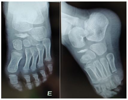
Kohler’s disease is a rare condition and describes an avascular necrosis of navicular bone. A 4-years-old female child came to emergency department limping, with intermittent left foot pain, with a month of evolution, refusing to bear weight, without history of trauma, fever or any illness. There was no swelling or redness of the foot. Complete blood count, C-reactive protein and erythrocyte sedimentation rate were normal. The diagnosis was made with foot radiographs, showing flattening and sclerosis of navicular bone. The disease was explained to parents and patient was discharged home with a course of NSAID to decrease symptoms.
Keywords: Kohler’s disease; Avascular necrosis; Foot pain; Limp.
Citation: Gameiro D, Miraldo L, Carvalhais P. Kohler’s disease: Clinical case of a limping young girl. J Clin Images Med Case Rep. 2025; 6(3): 3500.
Introduction
Kohler’s disease is a rare condition and describes an osteochondrosis of navicular bone (avascular necrosis) [1]. Typically affect boys between 3 and 7 years old [2]. Diagnosis can be made with radiographs showing flattening, sclerosis and navicular’s fragmentation [3]. Treatment consists in rest, pain control and limited weight bearing [4]. The prognosis is good due to radial arrangement of navicular blood supply, with almost all patients having restoration of bone structure [5].
Case description
A 4-years-old female child came to emergency department limping, with intermittent left foot pain, with a month of evolution, refusing to bear weight. Mother denied trauma, fever or any illness. There was no swelling or redness of the foot. Complete blood count, C-reactive protein and erythrocyte sedimentation rate were normal. The diagnosis was made with foot radiographs, showing flattening and sclerosis of navicular bone. The disease was explained to parents and patient was discharged home with a course of NSAID to decrease symptoms.
Conclusion
Although rare, this condition could be a cause of foot pain in children, even in female patients. A simple foot X-ray could make the diagnosis.
References
- Shastri N, Olson L, Fowler M. Kohler’s disease. Western Journal of Emergency Medicine. 2012; 13(1): 119-120.
- Shanley J, James DR, Lyttle M, Andronikou S, Knight DMA. Kohler’s disease: An unusual cause for a limping child. Arch Dis Child. 2017; 102(1): 101.
- Tuthill HL, Finkelstein ER, Sanchez AM, Clifford PD, Subhawong TK, et al. Imaging of Tarsal Navicular Disorders: A Pictorial Review. Foot Ankle Spec. 2014; 7(3): 210-224.
- Alhamdani M, Kelly C. Kohler’s disease presenting as acute foot injury. American Journal of Emergency Medicine. 2017; 35(11): 1787.e5-1787.e6.
- Deshpande S V, Channawar RA, Wamborikar H, Patil B, Pundkar A. Bilateral Kohler’s Disease: A Case Report. Cureus. 2023; 15(9): e44929.