
Journal of Clinical Images and Medical Case Reports
ISSN 2766-7820
Case Report - Open Access, Volume 6
Rehabilitation including neural gliding and exercises in the treatment of a “stinger” in a high school football player: A case study
Andy Waldhelm1,2*; Mareli Klopper3; Stephanie M Flynn1,4
1School of Rehabilitation, South College, 400 Goody Lane, Knoxville, TN, 37922, USA.
2Department of Physical Therapy, University of South Alabama, 5121 USA Dr. North, Mobile, AL 36688, USA.
3Department of Physical Therapy, Graceland University, 1401 W. Truman Rd. Independence, MO 6405, USA.
4Health Motion Physical Therapy, 3826 44th Street SE. Grand Rapids, MI 49512, USA.
*Corresponding Author : Andy Waldhelm
School of Rehabilitation, South College, 400 Goody
Lane, Knoxville, TN 37922, USA.
Email: awaldhelm@south.edu
Received : Feb 02, 2025
Accepted : Mar 03, 2025
Published : Mar 10, 2025
Archived : www.jcimcr.org
Copyright : © Waldhelm A (2025).
Abstract
Purpose: To describe the rehabilitation, including the use of neural gliding, on pain, impaired range of motion, strength, and function of a high school American football player suffering from a stinger injury.
Case description: The patient was a 17-year-old male who sustained a stinger while making a tackle. Initial symptoms included neck pain, shooting pain in his right upper extremity, and paresthesia over the lateral aspect of his right hand. The athlete complained of minimal neck soreness and shooting pain in right upper extremity, which he rated as a 9 out of 10 at worst. Objective findings included cervical right side flexion Active Range of Motion (AROM) limited by 50%, and right shoulder/scapular muscle strength 4 out of 5. Other findings included a positive Spurling’s test and upper limb neurodynamic test biasing the Median Nerve on the right where elbow extension provoked symptoms at 70 degrees of elbow flexion compared to 10 degrees on the left. Upper Extremity Functional Scale (UEFS) score was 71 out of 80. Physical therapy interventions included manual cervical distraction and joint mobilization, neural gliding interventions, strengthening of the shoulder complex and return to sport activities.
Outcomes: Upon discharge, the patient complained of intermittent neck pain of 2 out of 10 at worst, with no radicular symptoms. His UEFS score was 76 out of 80, AROM and strength were full, and all special tests were negative. The patient made a full return after two weeks and did not suffer another stinger during the remaining football season.
Discussion: There is limited research into using neural mobilization as an intervention to treat stingers. This case study describes the successful treatment and return to sport of a high school football player with recurrent stingers using neural gliding, but more research is warranted.
Keywords: Football; Stingers; Neural dynamics; Neural gliding; Manual therapy.
Citation: Waldhelm A, Klopper M, Flynn SM. Rehabilitation including neural gliding and exercises in the treatment of a “stinger” in a high school football player: A case study. J Clin Images Med Case Rep. 2025; 6(3): 3501.
Introduction
Transient brachial plexopathy, commonly referred to as a stinger or burner, is a frequent injury in collision sports such as rugby and American football [1-3]. A stinger typically involves injury to the cervical nerve roots or the brachial plexus, resulting in temporary sensory and/or motor deficits in one upper extremity, lasting from seconds to hours [4]. Although many stinger injuries go unreported, it is estimated that around half of all high school, college, and professional athletes will experience at least one stinger during their football careers [5,6], with a recurrence rate potentially as high as 87% [7].
The injury mechanism often involves a direct impact to the shoulder, causing contralateral cervical side-bending and concurrent ipsilateral shoulder girdle depression, which leads to a traction injury to the neurovascular bundle. Alternative mechanisms include compression of the nerve roots due to cervical spine hyperextension and ipsilateral side-bending or a direct blow to the brachial plexus at Erb’s point [2,4,8]. Notably, a significant proportion of stinger injuries in American football occur during tackles initiated by defensive linemen or linebackers [3].
Symptoms generally start with a shooting pain or burning sensation radiating from the neck down one upper extremity, followed by potentially prolonged impairment of sensory and motor function [9,10]. A study of stingers in young rugby players found that while nearly 80% of players experienced minimal time loss (0-1 day), neurological deficits could sometimes persist beyond one month [11].
Stinger injuries are diagnosed through a combination of history, physical examination, and diagnostic testing. The history should detail the mechanism of injury and all associated symptoms [12]. The physical examination should include an assessment of posture, the cervical spine, an upper quarter neurological examination, and palpation of key structures [2,12-14].
If symptoms persist beyond 24 hours or worsen, diagnostic imaging may be necessary. This may involve plain radiographs, Magnetic Resonance Imaging (MRI), Computed Tomography (CT), and/or a myelogram of the cervical spine and brachial plexus to identify the involved structures [2,10,14]. In cases where weakness persists, Electromyography (EMG) and nerve conduction studies, typically performed two to four weeks after the injury, can help differentiate between cervical radiculopathy and brachial plexus injury [15].
Managing a stinger injury can be challenging due to its variable healing times, and treatment should be tailored to the individual patient. A typical rehabilitation program for stinger injury recovery consists of three stages [2,12].
Stage 1) Rest, protection, and pain regulation: This stage focuses on managing pain and protecting the injured area. Physical modalities and medication may be used for pain relief [12,13].
Stage 2) Range of Motion (ROM), strengthening, flexibility, and postural correction: Therapeutic exercises and manual therapy are central during this stage. The focus is on regaining ROM, flexibility, strength, and scapular stability [2,12,16]. Postural re-education addresses issues such as forward head posture and rounded shoulders, which can increase pressure on the cervical nerve roots and brachial plexus [2,8,12,14].
Stage 3) Return to play progression: This stage begins only when the athlete has demonstrated full, pain-free active and passive ROM of the cervical spine and upper extremity, with no strength deficits and a normal neurological examination [2,10]. The return to play involves a gradual reintegration, starting with non-contact drills, progressing to controlled-contact activities, and eventually advancing to full-contact training [2].
Preventing initial and recurrent stinger injuries is crucial in contact sports such as football and rugby. Prevention strategies can be broadly categorized into three key areas: 1) tackling technique education, 2) preventative exercises, and 3) equipment fit and modification, though the evidence for the effectiveness of preventative exercises is limited [2,5,12,13].
An effective head and neck injury prevention program should focus on instructing or re-educating players on proper tackling techniques. Emphasis should be placed on avoiding leading with the head or helmet (spearing) and not dropping the shoulder during a tackle [2].
Although the evidence supporting preventative exercises is limited, these exercises are similar to those used in the rehabilitation of a stinger injury. They should aim to improve postural deviations, ROM restrictions, neck and shoulder weakness, and motor control/coordination. The goal is to help athletes better control the deceleration of their head, neck, and shoulders during play [2,5].
Properly fitting helmets and shoulder pads are essential for injury prevention. Additional equipment such as cervical collars, neck rolls, and shoulder pad lifters can help reduce movement or compression of the cervical spine and brachial plexus [2,8].
Symptoms of stinger injuries suggest neurological dysfunction, including shooting pain, burning sensations, sensory impairments, and muscle weakness. Although literature on this specific topic is limited, similar neurogenic symptoms have been successfully treated with neural mobilization in cases of upper quarter injuries such as cervical radiculopathy, cervicobrachial neurogenic pain, lateral epicondylitis, and adhesive capsulitis [17-20]. A systematic review and meta-analysis on the effectiveness of neural tissue management for nerve-related chronic musculoskeletal neck and arm pain concluded that neural tissue management is superior to minimal interventions (such as general exercise, mechanical traction, ultrasound, and joint mobilization) for pain relief and reducing disability [21]. The goal of neural mobilization is to improve the interaction between neural components and surrounding tissues by enhancing the nerve’s environment [22].
To date, no evidence specifically investigates the use of neural mobilization for treating stinger injuries. Therefore, the purpose of this case study is to describe the rehabilitation process, including the application of neural gliding techniques, and its impact on pain, range of motion, strength, and function in a high school American football player suffering from a stinger injury.
Case description
The patient was a 17-year-old male high school senior who sustained an injury to his neck and right upper extremity during a tackle while playing defensive end in his American football team’s first game of the season. He was assessed immediately by the athletic trainer, physical therapist, and team physician. Following these evaluations, the athlete did not return to play in that game. The next day, he followed up with the team physician and was subsequently referred to physical therapy.
Three days after the injury, the same physical therapist conducted a thorough evaluation. The patient had a prior history of a stinger injury to the same side during the final game of the previous football season. He had managed to return to play that night and reported no symptoms during the off-season, receiving no treatment for the previous injury. The patient had no other significant medical history and was cleared for red flags, including headache, nausea, loss of memory, dizziness, and difficulty swallowing.
Examination
The patient reported an immediate onset of neck pain and shooting pain radiating into his right upper extremity, accompanied by paresthesia over the lateral aspect of his right hand following the injury. During the physical therapy evaluation, he described minimal soreness in his neck, rated as 2 out of 10 on the Numerical Pain Rating Scale (NPRS). However, he continued to experience significant shooting pain in his right upper extremity, with a peak intensity of 9 out of 10 on the NPRS.
(Table 1) lists a full objective examination with key objective findings:
● Cervical spine side-bending to the right (AROM) limited by 50%
● Gross muscle strength of the right shoulder/scapular musculature measured at 4 out of 5
● Positive Spurling’s test on the right with an increase in radicular pain in right upper extremity
● Positive cervical distraction tests with a reduction in radicular symptoms
● Positive cervical distraction tests with a reduction in radicular symptoms
● Upper Extremity Functional Index (UEFI) score of 71 out of 80 where a score of 80 out to 80 is equal to highest functional status.
Clinical impression
The patient presented with findings consistent with a recurrent stinger injury on the right, resulting from a traction injury to the brachial plexus sustained during a tackle. He exhibited cervical spine Range of Motion (ROM) impairments, weakness in the right shoulder and scapular musculature, and positive neurodynamic findings on the right.
After ruling out red flag conditions, a differential diagnosis between cervical radiculopathy and stinger injury was further assessed using a cluster of tests: upper limb neurodynamics, Spurling’s test, cervical distraction, and cervical rotation Active Range of Motion (AROM). With cervical rotation AROM greater than 60 degrees bilaterally, the positive likelihood ratio (+LR) for cervical radiculopathy decreased from 30.3 to 6.1 [23-25]. The mechanism of injury, the patient’s age, the absence of a dermatomal pain pattern, and more pronounced motor symptoms compared to sensory deficits supported the conclusion that the patient was more likely experiencing a stinger injury rather than cervical radiculopathy [24,26].
Table 1: Examination findings at initial evaluation and discharge.
| Initial evaluation | Discharge (12 visits) | ||
|---|---|---|---|
| Active cervicalROM(goniometer) | Flexion | 40 deg | 55 deg |
| Extension | 35 deg | 50 deg | |
| Right Rotation | 70 deg | 80 deg | |
| Left Rotation | 80 deg | 85 deg | |
| Right SB | 20 deg | 35 deg | |
| Left SB | 40 deg | 40 deg | |
| Active shoulderelevation ROM (goniometer) | Right | 160 deg | 175 |
| Left | 175 deg | 175 | |
| Rightupper extremity strength (out of 5) | Shoulder flexion | 4- | 5 |
| Shoulder abduction | 4 | 5 | |
| Shoulder ER | 4- | 5- | |
| Shoulder IR | 4+ | 5 | |
| Middle Trapezius | 4 | 5 | |
| Lower Trapezius | 4- | 4+ | |
| C2-6 myotome | Strong | Strong | |
| C7,8 myotome | Weak andpain free | Strong | |
| Bicep and triceps DTR | 2+ | 2+ | |
| Joint accessorymobility | Upper C/Sright UPA | Hypomobile/ painful | Normal/painfree |
| Lower C/S right UPA | Normal/ painful | Normal/painfree | |
| Sensation | C8 dermatome | Impaired tolight touch | Normal |
| Special tests | Spurling | Reproduce symptomson right | Negative |
| Cervical Distraction | Decrease in radicular symptoms | N/T due to lack of radicular symptoms | |
| Right ULNTMedian | Reproduce symptomsat 70 deg ofelbow flexion | Symptoms not reproduced and ableto achieve 10 deg of elbowflexion | |
| Right ULNTUlnar | Negative | N/T | |
| C/S flexion/rotationtest | Right: 20 degLeft: 40 deg | Right: 40 deg Left: N/T | |
| Brachial plexus compression tests | Reproduce shooting pain inright arm | Negative |
Note: Negative test: Did not reproduce symptoms; ROM: Range of Motion; SB: Side Bending; deg: degrees; C/S: Cervical Spine; DTR: Deep Tendon Reflex; UPA: Unilateral Posterior to Anterior glides; ULNT: Upper Limb Neurodynamic Test; N/T: Not Tested.
Further classification of the severity of the nerve injury was made using the Seddon-based classification for peripheral nerve injuries. Grade I stingers, the present case, classified as neuropraxia, involve minimal structural damage and are expected to recover fully within a short period. These typically present with pain, muscular weakness, and numbness, but lack muscular atrophy [27,28]. Grade II stingers, or axonotmesis, also result in full recovery but require a longer healing time due to damage to the axon and myelin while preserving the internal nerve structures. Patients with Grade II stingers may present with pain, muscle wasting, and both motor and sensory loss [27,28]. Grade III injuries, classified as neurotmesis, represent the most severe form and are rare in this type of injury. Neurotmesis is characterized by muscle atrophy and complete motor and sensory loss due to extensive nerve damage [27,28].
Outcome measure
The Upper Extremity Functional Index was used to assess the patient’s primary complaints, which were localized to his right upper extremity rather than the cervical or upper thoracic spine. The patient’s initial score was 71 out of 80, with lifting, carrying, and throwing a ball being the main functional activity deficits.
Interventions
The patient was seen in physical therapy for 2 to 3 visits a week for five weeks, 12 visits in total. A summary and progression of the interventions are listed in (Table 2). The goals of Week 1 were to improve cervical spine range of motion, neural mobility of the median nerve, and activation/muscular endurance of the scapular stabilizing muscles. Manual therapy, including sustained cervical traction into resistance, right-sided Unilateral Posterior to Anterior (UPA) grades III and IV mobilization, and pain-free cervical retraction and extension active range of motion exercises were implemented to improve mobility of the cervical spine.
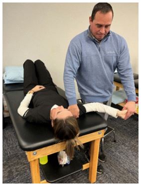
Neural mobilization was introduced as pain-free glider (Figure 1). In a supine position, the physical therapist passively positioned the patient’s right shoulder in 90 degrees of abduction and maximal external rotation. The patient’s fingers were extended, and his right wrist was maximally extended and supinated before the therapist applied passive right elbow extension within a pain-free ROM. Gliders were performed as pain-free PROM/AAROM by repeatedly flexing and extending the patient’s elbow. These techniques increased pain-free ROM during elbow extension and decreased paresthesia in the right hand.
To maintain and further capitalize on the gains made during physical therapy sessions using neural gliders, the patient was instructed in a home exercise program to replicate the neural slider. Neural gliding exercises were performed in standing and involved replicating the upper extremity movements performed in a pain-free elbow extension ROM. The patient was instructed to raise his right arm to 90 degrees of shoulder abduction, followed by maximal external rotation. After extending his fingers, maximally extending his wrist, and supinating his forearm, the patient extends his elbow to the onset of his familiar symptoms before returning to a position of elbow flexion. The patient performed the neural gliding exercises in two sets of fifteen repetitions, three times a day.
Therapeutic exercises, including standing rows and I, T, Y exercises, were initially performed with no resistance and then progressed to light resistance to increase muscle activation and muscle endurance of the rhomboids, middle and lower trapezius muscles. The patient performed each exercise for two sets of fifteen repetitions. Following the first week of physical therapy, the athlete had significant improvements in cervical ROM and neural mobility, shown by an increase in elbow extension when performing the Median Nerve upper limb neurodynamic test.
The goals of Week 2 were to achieve full cervical range of motion and increase the strength and stability of the shoulder complex so the athlete would be cleared to return to all football activities during his follow-up visit with his physician, which occurred at the end of Week 2. Manual therapy interventions were minimal and included grade IV right UPA and right side glide mobilization to the lower cervical spine to obtain the last few degrees of right cervical side bend. Cervical AROM exercises and nerve gliding were reviewed during the first session of Week 2 and then allocated to the home exercise program.
Upper quarter stabilization and strengthening became the focus of Week 2 as several new exercises were included in the rehabilitation program. Dynamic Bodyblade (Hymanson, Inc. Playa del Rey, CA USA) exercises in the sagittal, frontal, and transverse planes were included in the dynamic warm-up to activate the muscles of the shoulder complex before resistance training. Progressive resistance training used elastic bands and dumbbells, and the exercise parameters used included three to four sets, six to eight reps with one to two minutes of rest between sets and exercises. Resistance for the row, full can, I, T, Y exercises increased from Week 1 into Week 2, and new resistance exercises included shoulder internal and external rotation at 0 degrees of abduction, lat pull-downs to strengthen the latissimus dorsi muscle, and wall push-up plus was used to strengthen the serratus anterior muscle in a closed-chain position. Isometric neck strengthening exercises were also included to improve neck strength and function. Following Week 2, the athlete was cleared to return to all football activities after regaining full neck AROM, improved shoulder strength and stability, and eliminating radicular symptoms in his right shoulder.
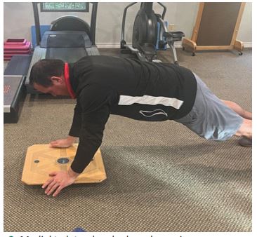
Although the athlete was cleared to return to sport, physical therapy was continued for three more weeks. Weeks 3-5 goals were to continue improving shoulder strength and stability and progress to plyometric and sport-specific activities. During Weeks 3-5 of the rehabilitation program, no manual therapy was performed, but the athlete continued to perform the neural gliding exercise independently. Plyometrics became one of the two main components of the last three weeks of rehabilita tion, with new exercises such as single arm medicine ball drops at different shoulder angles, chest and overhead pass/catch, and plyometric push-ups included in the program. A new closed chain dynamic stability exercise, upper extremity rocker board in the anterior/posterior and medial/lateral directions, was also introduced (Figure 2).
Table 2: Week-by-week progression of physical therapy interventions.
| Initial evaluation | Discharge (12 visits) | ||
|---|---|---|---|
| Active cervical ROM (goniometer) | Flexion | 40 deg | 55 deg |
| Extension | 35 deg | 50 deg | |
| Right Rotation | 70 deg | 80 deg | |
| Left Rotation | 80 deg | 85 deg | |
| Right SB | 20 deg | 35 deg | |
| Left SB | 40 deg | 40 deg | |
| Active shoulder elevation ROM (goniometer) | Right | 160 deg | 175 |
| Left | 175 deg | 175 | |
| Right upper extremity strength (out of 5) | Shoulder flexion | 4- | 5 |
| Shoulder abduction | 4 | 5 | |
| Shoulder ER | 4- | 5- | |
| Shoulder IR | 4+ | 5 | |
| Middle Trapezius | 4 | 5 | |
| Lower Trapezius | 4- | 4+ | |
| C2-6 myotome | Strong | Strong | |
| C7,8 myotome | Weak and pain free | Strong | |
| Bicep and triceps DTR | 2+ | 2+ | |
| Joint accessory mobility | Upper C/S right UPA | Hypomobile/ painful | Normal/pain free |
| Lower C/S right UPA | Normal/ painful | Normal/pain free | |
| Sensation | C8 dermatome | Impaired to light touch | Normal |
| Special tests | Spurling | Reproduce symptoms on right | Negative |
| Cervical Distraction | Decrease in radicular symptoms | N/T due to lack of radicular symptoms | |
| Right ULNT Median | Reproduce symptoms at 70 deg of elbow flexion | Symptoms not reproduced and able to achieve 10 deg of elbow flexion | |
| Right ULNT Ulnar | Negative | N/T | |
| C/S flexion/rotation test | Right: 20 deg Left: 40 deg | Right: 40 deg Left: N/T | |
| Brachial plexus compression tests | Reproduce shooting pain in right arm | Negative |
Note: UPA: Unilateral Posterior to Anterior mobilization; C/S: Cervical Spine; 90/90: upper extremity positioned at 90 degrees of shoulder abduction and 90 degrees of elbow extension; ER: External Rotation; IR: Internal Rotation
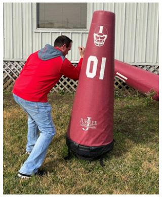
The second component introduced was the return to sport activities. Since the athlete was a defensive lineman, positionspecific activities were performed to ensure he was safe to return to sport. Activities included acceleration from a threepoint stance, pushing a sled, engaging and getting off of a block from an offensive lineman, and performing a swim or rip move against a large blocking dummy (Figure 3). Resistance training continued with a progression that included shoulder internal and external rotation at 90 degrees of shoulder abduction and elbow flexion, and push-up plus from the floor. There was also an adjustment in exercise parameters in Weeks 3-5 to focus on strength and power; they included: Three to five sets, four to six repetitions with two minutes of rest between sets and exercises. The athlete was able to play a full game at the end of Week 3 of his rehabilitation with minimal neck soreness and no radicular symptoms.
Outcomes
Upon discharge from physical therapy, the patient complained of intermittent neck pain of 2 out of 10 at worst, without radicular symptoms in his right upper extremity. Key objective findings at discharge were as follows (Table 1):
● Cervical spine AROM was full and pain-free in all directions
● Gross muscle strength of shoulder/scapular musculature at 5 out of 5
● Spurling’s test was negative
● Median nerve upper limb neurodynamic test was negative with elbow extension equal between the left and right extremities and no reproduction of symptoms
● Upper Extremity Functional Scale (UEFS) score of 76 out of 80
The patient made a full return to football two weeks after the injury and did not suffer symptoms or another stinger during the rest of the football season.
Discussion
There is currently limited evidence regarding the use of neurodynamic mobilization techniques in managing stinger injuries. This case study highlights the incorporation of neural mobilization techniques to address an athlete’s complaints of neck and arm pain, along with paresthesia, following successive stinger injuries.
Neural mobilization is a movement-based therapy frequently utilized by physical therapists to address nerve-related symptoms. Studies in both humans and animals have shown that neurodynamic mobilization techniques can decrease intraneural edema, improve intraneural fluid dispersion, reduce thermal and mechanical hyperalgesia, and reverse the heightened immune response following a nerve injury [22].
In the case of the 17-year-old athlete, the decision to incorporate neurodynamic interventions was based on symptoms indicative of neural pathology, such as numbness, tingling, and weakness. Within a week of starting therapy, the patient reported a reduction in numbness and tingling, and showed improved strength, cervical spine AROM, and neurodynamic mobility.
While we can only speculate about the physiological mechanisms behind his positive response, it is possible that the neurodynamic sliders used during treatment helped disperse intraneural edema and reduce the immune response associated with his injury.
The suspected mechanism of injury influences the clinician’s choice of neurodynamic intervention. Two common techniques are employed in clinical practice: Tensioners, which aim to lengthen the nerve, and sliders (also known as gliders or flossing), which create tension on one end of the nerve bed while releasing it at the other. Studies have shown that sliders facilitate greater neural excursion with less strain compared to tensioners [29,30].
When treating a patient with a stinger injury caused by a traction mechanism, it is preferable to start with sliders. This approach impacts neural movement without exacerbating strain or replicating the traction injury mechanism in a sensitized nerve. Continuous reassessment and adaptation of the neurodynamic intervention are essential based on the patient’s evolving presentation. If pain decreases and neural excursion improves, it may then be appropriate to introduce tensioning techniques.
To the authors’ knowledge, there is limited research on the impact of neural mobilization on athletic injury recovery. Most existing studies emphasize its benefits for functional flexibility, demonstrating a correlation between improved flexibility and neural mobilizations [31]. For example, research involving a group of Australian football players with grade one hamstring injuries showed that those treated with neural gliding in conjunction with traditional treatment experienced faster recovery and return to play compared to those receiving traditional treatment alone [32]. Additionally, a case study of an elite baseball pitcher with posterior interosseous neuropathy found that neural mobilizations combined with conservative rehabilitation allowed the athlete to return to play 38 days post-injury, achieving baseline velocity with no reported symptoms [33]. While the literature on neural mobilization in athletic injury recovery is limited, positive outcomes suggest its potential benefits in facilitating successful returns to play after injury.
Unfortunately, the muscle testing conducted during this patient’s examination did not include the use of a dynamometer. A hand-held dynamometer is user-friendly and would have provided valuable baseline strength data, as well as an objective measure of strength changes. Additionally, grip strength testing, a potentially significant indicator of strength changes, was not performed. Lastly, the athlete was cleared to play by the physician before a return-to-sport assessment could be conducted. Ideally, both the shot-put test and closed kinetic chain upper extremity tests should be utilized to evaluate an athlete’s readiness to return to sport.
This case report details the successful treatment of a patient with a stinger injury using neurodynamic interventions as part of a comprehensive treatment plan. While we cannot imply causation or generalize from a single case, the limited literature guiding treatment for this population underscores the need for further research. A case series would strengthen the evidence base and help clinicians optimize care for these patients.
Conclusion
This case study suggests that incorporating neural gliding into a comprehensive rehabilitation program can effectively restore range of motion, strength, and overall function in a high school football player with recurring stinger injuries. After five weeks of using passive and active median nerve glides as part of his rehabilitation, the athlete successfully returned to play with pre-injury strength and mobility. Thus, neural glides may be a valuable addition to treatment protocols for stinger injuries; however, further research is warranted to better understand their efficacy.
References
- Meyer SA, Schulte KR, Callaghan JJ, et al. Cervical spinal stenosis and stingers in collegiate football players. Am J Sport Med. 1994; 22(2): 158-166.
- Feinberg JH. Burners and stingers. Phys Med Rehabil Clin N Am. 2000; 11(4): 771-784.
- Green J, Zuckerman SL, Dalton SL, et al. A 6-year surveillance study of Stingers in NCAA American Football. Res Sports Med. 2017; 25(1): 26-36.
- Ahearn, Briggs M, Starr, Harlan M, Seiler, John G. Traumatic Brachial Plexopathy in Athletes: Current Concepts for Diagnosis and Management of Stingers. J Am Acad Orthop Surg. 2019; 27(18): 677-684.
- Cramer CR. A reconditioning program to lower the recurrence rate of brachial plexus neurapraxia in collegiate football players. J Athl Train. 1999; 34(4): 390-396.
- Starr HM Jr, Anderson B, Courson R, Seiler JG. Brachial plexus injury: A descriptive study of American football. J Surg Orthop Adv. 2014; 23(2): 90-7.
- Sallis RE, Jones K, Knopp W. Burners: Offensive strategy for an under-reported injury. Physician Sportmed. 1992; 20(11): 47-55.
- Shannon B. Klimkiewicz JJ. Cervical burners in the athlete. Clin Sports Med. 2002; 21(1): 29-35.
- Levitz CL, Reilly PJ, Torg JS. The pathomechanics of chronic, recurrent cervical nerve root neurapraxia. Am J Sport Med. 1997; 25(1): 73-76.
- Standaert CJ, Herring SA. Expert opinion and controversies in musculoskeletal and sports medicine: Stingers. Arch Phys Med Rehabil. 2009; 90: 402-406.
- Kawasaki T, Ota C, Yoneda T, et al. Incidence of Stingers in Young Rugby Players. Am J Sports Med. 2015; 43(11): 2809-15.
- Weinstein SM. Assessment and rehabilitation of the athlete with a “Stinger”: A model for the management of non-catastrophic athletic cervical spine injury. Clin Sports Med. 1998; 17(1): 127- 135.
- Weinberg J, Rokito S, Silber JS. Etiology, treatment, and prevention of athletic stingers. Clin Sports Med. 2003; 21: 493-500.
- Tosti R, Rossy W, Sanchez A, Lee SG. Burners, Stingers, and other brachial plexus injuries in the contact athlete. Oper Tech Sports Med. 2016; 24: 273-277.
- Bowles DR, Canseco JA, Alexander TD, Schroeder GD, Hecht AC, et al. The Prevalence and Management of Stingers in College and Professional Collision Athletes. Curr Rev Musculoskelet Med. 2020; 13(6): 651-662.
- Hartley RA, Kordecki ME. Rehabilitation of chronic brachial plexus neuropraxia and loss of cervical extension in a high school football player: A case report. Int J Sports Phys Ther. 2018; 13(6): 1061-1072.
- Savva C, Giakas G. The effect of cervical traction combined with neural mobilization on pain and disability in cervical radiculopathy. A case report. Man Ther. 2013; 18: 443-446.
- Coppieters MW, Stappaerts KH, Wouters LL, Janssens K. The immediate effects of a cervical lateral glide treatment technique in patients with neurogenic cervicobrachial pain. J Orthop Sports Phys Ther. 2003; 33: 369-378.
- Vincenzio B, Collins D, Wright A. The initial effects of a cervical spine manipulative physiotherapy treatment on pain and dysfunction of lateral epicondylalgia. Pain. 1996; 68(1): 69-74.
- Farrell K, Lampe K. Addressing neurodynamic irritability in a patient with adhesive capsulitis: A case report. J Man Manip Ther. 2017; 25(1): 47-56.
- Su Y, Lim EC. Does Evidence Support the Use of Neural Tissue Management to Reduce Pain and Disability in Nerve-related Chronic Musculoskeletal Pain? A Systematic Review With MetaAnalysis. Clin J Pain. 2016; 32(11): 991-1004.
- Basson A, Olivier B, Ellis R, Coppieters M, Stewart A, et al. The effectiveness of neural mobilization for neuromusculoskeletal conditions: A systematic review and meta-analysis. J Orthop Sports Phys Ther. 2017; 47(9): 593-615.
- Wainner RS, Fritz JM, Irrgang JJ, Boninger ML, Delitto A, Allison S. Reliability and diagnostic accuracy of the clinical examination and patient self-report measures for cervical radiculopathy. Spine (Phila Pa 1976). 2003; 28(1): 52-62.
- Yarznbowicz R, Wlodarski M, Dolutan J. Classification by pain pattern for patients with cervical spine radiculopathy. J Man Manip Ther. 2020; 28(3): 160-169.
- Waldrop MA. Diagnosis and treatment of cervical radiculopathy using a clinical prediction rule and a multimodal intervention approach: a case series. J Orthop Sports Phys Ther. 2006; 36(3): 152-9.
- Elias I, Pahl MA, Zoga AC, Goins ML, Vaccaro AR. Recurrent burner syndrome due to presumed cervical spine osteoblastoma in a collision sport athlete-a case report. Journal of brachial plexus and peripheral nerve injury. 2007; 2(1): 13.
- Seddon HJ. A Classification of Nerve Injuries. Br Med J. 1942; 2(4260): 237-239.
- Kuhlman GS, McKeag DB. The burner: A common nerve injury in contact sports. Am Fam Physician. 1999; 60(7): 2035-2042.
- Coppieters MW, Butler DS. Do ‘sliders’ slide and ‘tensioners’ tension? An analysis of neurodynamic techniques and considerations regarding their application. Man Ther. 2008; 13(3): 213- 221.
- Coppieters MW, Andersen LS, Johansen R, et al. Exercises: An in vivo cross-sectional study using dynamic ultrasound imaging. J Orthop Sports Phys Ther. 2015; 45(10): 731-737.
- Balcı A, Ünüvar E, Akınoğlu B, Kocahan T. The effect of different neural mobilization exercises on hamstring flexibility and functional flexibility in wrestlers. J Exerc Rehabil. 2020; 16(6): 503- 509.
- Kornberg C, Lew P. The effect of stretching neural structures on grade one hamstring injuries. J Orthop Sports Phys Ther. 1989; 10(12): 481-487.
- Robb A, Sajko S. Conservative management of posterior interosseous neuropathy in an elite baseball pitcher’s return to play: A case report and review of the literature. J Can Chiropr Assoc. 2009; 53(4): 300-310.