
Journal of Clinical Images and Medical Case Reports
ISSN 2766-7820
Clinical Image - Open Access, Volume 6
Parafoveal metallic fragment after pars plana vitrectomy
Vincent Huang*; Amro Omari; Benjamin J Kim
Department of Ophthalmology, Scheie Eye Institute, University of Pennsylvania, Philadelphia, PA, USA.
*Corresponding Author : Vincent Huang
Department of Ophthalmology, Scheie Eye Institute,
University of Pennsylvania, Philadelphia, PA, USA.
Email: vincent.huang@pennmedicine.upenn.edu
Received : Feb 03, 2025
Accepted : Mar 04, 2025
Published : Mar 11, 2025
Archived : www.jcimcr.org
Copyright : © Huang V (2025).
Citation: Huang V, Omari A, Kim BJ. Parafoveal metallic fragment after pars plana vitrectomy. J Clin Images Med Case Rep. 2025; 6(3): 3504.
Description
A 71 year-old male underwent right eye retinal detachment repair with pars plana vitrectomy and C3F8 gas. Three months later, the visual acuity was 20/25, and the retina was attached. However, the patient mildly complained of a “glint” in his right eye vision when in bright light. Evaluation revealed a tiny refractile deposit nasal to the fovea (Figure 1A) that was not present on pre-operative exam. The deposit was hyper-reflective on OCT (Figure 1B) and located on the nerve fiber layer. The option of foreign body surgical removal was discussed, but he elected to monitor given his good visual outcome.
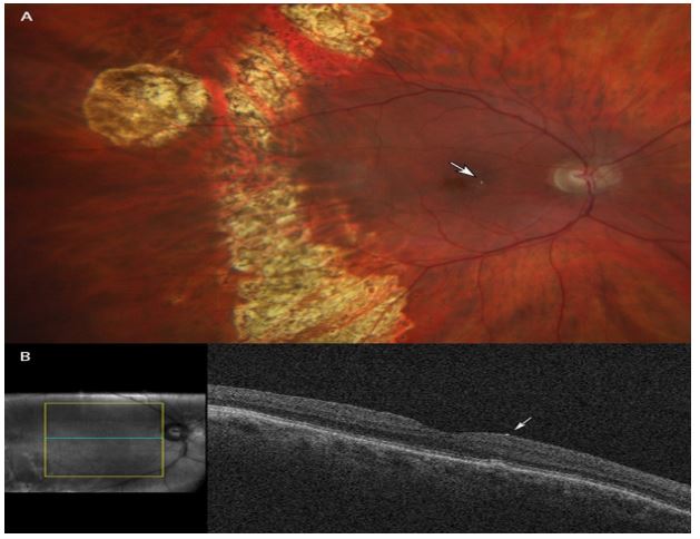
(B) OCT demonstrating preretinal hyperreflective particle (arrow) nasal to the fovea.