
Journal of Clinical Images and Medical Case Reports
ISSN 2766-7820
Case Report - Open Access, Volume 6
A newborn presenting with clinical cholestasis in the first hour of life
Imen Ebdelli*; Farida Friha; Asma Marzouk; Ahlem Kefi; Rahma Thabti; Asma Bouaziz
Pediatrics Department, Ben Arous Regional Hospital, Tunisia.
*Corresponding Author : Imen Ebdelli
Pediatrics Department, Ben Arous Regional Hospital,
Tunisia.
Email: imen.ebdeli@gmail.com
Received : Feb 10, 2025
Accepted : Mar 07, 2025
Published : Mar 14, 2025
Archived : www.jcimcr.org
Copyright : © Ebdelli I (2025).
Citation: Ebdelli I. A newborn presenting with clinical cholestasis in the first hour of life. J Clin Images Med Case Rep. 2025; 6(3): 3509.
Introduction
Neonatal cholestasis is a dysfunction of hepatic cells and/ or capillary bile secretion in newborns, leading to impaired bile flow and the accumulation of fatty acids, bilirubin, and other substances in the liver and bloodstream. Typically observed within the first two weeks of life, neonatal cholestasis can be caused by both congenital and acquired viral infections. Among these, the herpesvirus group, especially Cytomegalovirus (CMV), has been identified as a significant cause of intrahepatic neonatal cholestasis [2]. Previous studies, based on serological testing, indicate that CMV plays a role in the development of neonatal cholestasis, with rates of congenital infection ranging from 0.4% to 1.0% in the United States, and up to 2% in some regions of Asia and Africa [3]. The hematogenous transmission rate of CMV is between 0.5% and 2.5%. Early maternal-fetal transmission worsens prognosis and increases the risk of severe anomalies [4]. The transmission rate of CMV to the fetus is about 30% during primary infection, while it is lower (1-2%) in cases of non-primary infection. Although primary infection carries an increased risk, most congenital infections are nonprimary, particularly in regions with high immunization rates, such as Africa, South America, and Asia.
Infection may be asymptomatic in 90% of cases, with affected newborns being detected mainly through neonatal screening programs. However, 10% of infected newborns exhibit symptoms, including hepatosplenomegaly, jaundice, and neurological abnormalities [3]. This article presents a rare case of early cholestatic jaundice observed at one hour of life, associated with congenital CMV infection. It highlights the diagnostic challenges, clinical variability, and evolving nature of this condition.
Case presentation
Newborn AA, born from a non-consanguineous marriage The mother, a 21-year-old primiparous homemaker, was nonsmoker, non-alcoholic, had no significant medical history, and had no identifiable infectious history: no Premature Rupture of Membranes (PROM), no maternal fever, and no vaginal swabs or urinalysis performed. Her pregnancy was poorly monitored, with no prenatal serologies or ultrasounds beyond a third-trimester scan showing microcephaly. The baby was delivered at term (40 weeks + 3 days of amenorrhea) via spontaneous vaginal delivery at the Regional Hospital of Ben Arous, Tunisia, in the context of fetal distress (micro-oscillating fetal heart rate). At birth, the amniotic fluid was stained, and the newborn exhibited poor adaptation to extrauterine life, with an Apgar score of 5 at 1 minute and 7 at 5 minutes.
He was admitted to the neonatal unit immediately after birth for neonatal respiratory distress with severe symmetrical intrauterine growth restriction (birth weight: 2700 g, head circumference: 30 cm, length: ...). The respiratory distress improved quickly within 2 hours of life with supportive care (Silverman score of 2 at 10 minutes of life, then 0 at 2 hours). He presented with hypoglycemia at 0.2 g/dL, requiring correction with 0.3 cc/ kg/h of glucose infusion. At 1 hour of life, a greenish jaundice was noted along with purpuric spots initially on the face and extending to the trunk. The newborn displayed dysmorphic features, including retrognathia, a broad nose, and a prominent forehead. Examination revealed hepatosplenomegaly, axial hypotonia, a 3 cm cephalohematoma, and bilateral subconjunctival hemorrhages. The urine and meconium were normally colored.
Laboratory tests revealed thrombocytopenia, hepatic cholestasis, hepatic cytolysis without signs of hepatic failure, and evidence of asphyxia. The baby was started on simplified meningitis-dose antibiotics following a normal lumbar puncture.
Biliary atresia was suspected, and an emergency abdominal ultrasound was performed, which showed no abnormalities. A transfontanellar ultrasound, conducted due to the microcephaly, revealed quadriventricular hydrocephalus, enlargement of the cisterna magna, and partial agenesis of the corpus callosum. A subsequent brain CT scan indicated findings consistent with congenital CMV embryofetopathy, including diffuse periventricular and subcortical white matter calcifications, periaqueductal and cerebellar calcifications, bilateral bulbar calcifications, atrophy of the corpus callosum tail, diffuse cerebral and cerebellar atrophy with cortical sulci rarefaction, right parietal cephalhematoma (32 mm x 13 mm), and dysgenesis of the corpus callosum. Echocardiography revealed a patent foramen ovale (PFO) with left-to-right shunting and a small patent ductus arteriosus (PDA) with left-to-right shunting.
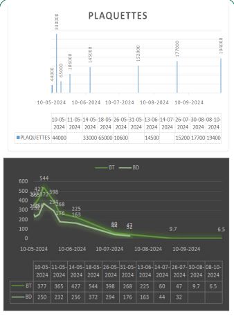
TORCH serology was performed, revealing positive CMV PCR in the urine with a viral load of 1,282,200,000 copies/mL. Antviral therapy with ganciclovir was indicated but unavailable in the hospital. The patient was managed symptomatically with ursodeoxycholic acid, vitamin supplements, and a cow’s milk hydrolysate-based diet. The evolution was marked by an improvement in cholestasis (Figure 1).
Discussion
Overall clinical, laboratory, and imaging results in the case in question are typical of a CMV congenital infection which is the most common congenital viral infection, occurring in 0.4-2.3% of all live births, and is probably a common cause of mental retardation and nonhereditary sensorineural deafness in children [5].
Common clinical signs include hepatosplenomegaly (60%), microcephaly (53%), jaundice (67%), petechiae (76%), and at least one neurological anomaly (68%). Laboratory abnormalities include increased transaminases (83%), thrombocytopenia (77%), direct hyperbilirubinemia (69%), hemolysis (51%), and hyperproteinorrachia (46%) [3]. Inguinal hernia and chorioretinitis are less common [5]. Intrauterine growth restriction has recently been identified as a sign of symptomatic congenital CMV infection by several authors [3]. In this case, the neonate presented microcephaly, hepatomegaly, and jaundice, with a notable increase in direct bilirubin probably due to cholestasis caused by CMV invasion of the intrahepatic bile duct epithelium.
Biliary atresia, idiopathic hepatitis, sepsis, alpha-1 antitrypsin deficiency, and galactosemia are among the common causes of infant cholestatic jaundice. Before making a diagnosis of CMV infection, these should be taken into account. Due to a lack of techniques for diagnosis and a lack of awareness about the condition, many cases of prenatal CMV infection remain undetected in poor nations [6].
In this particular case, microcephaly was discovered during a third-trimester prenatal obstetric ultrasound. Expectant attitudes were used when universal CMV screening was not available. Transfontanellar ultrasound and brain scan were used to confirm the diagnosis postnatally, and the results showed microcephaly, periventricular calcifications, and cerebral and cerebellar atrophy. Clinical professionals may plan for and expect a comprehensive assessment of the newborn by using prenatal data to undertake the diagnostic tests required to confirm congenital CMV infection. It should be mentioned that up to 50% of people infected with CMV do not exhibit any ultrasonography symptoms, making prompt diagnosis even more difficult [7]. Radiographic findings in the brain are abnormal in approximately 50%-70% of children with symptomatic infection at birth, with the most common finding being intraventricular calcification in 79% of patients [7].
We consider this case a primary congenital CMV infection, characterized by the appearance of jaundice in the first few hours of life which is a rare manifestation not found in the literature. In a Bangladeshi study, the mean age of onset was 12.4±2.8 days [8]. A case report in Nigeria showed Jaundice appeared on the third day of life [6].
In the index case, the diagnosis of congenital CMV infection was made based on elevated CMV Polymerase Chain Reaction (PCR) in urine which is considered as the gold standard for confirming congenital CMV infection. The recovery of replicating virus and/or viral nucleic acids within the first 2-3 weeks of life is required for the diagnosis of congenital CMV infections. Urine, saliva, and blood are all sources of the virus and viral nucleic acids. Routine viral culture paired with immunofluorescence and PCR is among the detection methods [3]. Several clinical trials have looked at the treatment of congenitally infected children with ganciclovir, and a significant number of infected infants have been treated off-label with this medicine for severe CMV infections [3]. Two studies funded by the NIH’s Collaborative Antiviral Study Group (CASG) suggest that 6 weeks of intravenous ganciclovir or 6 months of oral ganciclovir may help prevent hearing loss and improve developmental outcomes in infected newborns [3].
However, in our case, this treatment was not available, posing a real therapeutic challenge. Despite this limitation, the clinical course was marked by spontaneous resolution of the cholestasis with symptomatic treatment, including nutritional support and measures to prevent complications. This finding highlights the variability of the clinical presentation and course of congenital CMV infections and the importance of appropriate management even in the absence of specific antiviral treatment.
It should be emphasised that the unavailability of treatment in certain regions may limit therapeutic options and justifies the need to improve access to antivirals in countries with limited resources. Nevertheless, the overall evolution of the child’s condition was marked by repeated episodes of severe bronchiolitis requiring mechanical ventilation, which eventually led to his death. This case illustrates the complexity of congenital CMV infections, where recurrent respiratory complications can impair long-term prognosis, even when the initial liver outcome is favorable. It also highlights the need to strengthen access to specific treatments, particularly in resource-limited settings, as well as the importance of multidisciplinary follow-up to identify and manage secondary complications. This presentation raises a number of questions: did the patient have an increased susceptibility to recurrent respiratory infections due to lung damage associated with CMV infection? Was the lung pathologically impaired? Did this respiratory comorbidity increase the risk of mortality in this context?
These questions highlight the need for a better understanding of the interactions between congenital CMV infection and potential multisystem damage, particularly to the lungs. They also highlight the importance of prolonged follow-up and multidisciplinary management to detect and manage associated complications at an early stage, which can have a significant impact on long-term prognosis.
Conclusion
This case of neonatal cholestasis due to congenital CMV infection presents several important clinical lessons. The early onset of jaundice, observed in the first hour of life, is a rare manifestation of congenital CMV infection, which typically presents with symptoms after a few days. Despite the unavailability of antiviral treatment in our case, the neonate showed improvement with symptomatic management, highlighting the variability in the clinical course of congenital CMV infections.
The case also emphasizes the importance of early diagnosis and the challenges of managing congenital CMV in regions with limited resources. Although ganciclovir is considered the gold standard treatment, its unavailability in resource-poor settings underscores the need for better access to antiviral therapies. Furthermore, the recurrent respiratory complications in this case raise questions about the long-term impact of CMV-related multisystem damage, particularly lung involvement, and the need for comprehensive follow-up and multidisciplinary management.
This case highlights the complexity of congenital CMV infections and the necessity for continued research and improved management strategies to optimize outcomes for affected infants, particularly in low-resource settings.
Funding: none.
Conflict of interest: none.
References
- Liu P, Guo L, Huang L, et al. Analysis of factors affecting the prognosis of neonatal cholestasis.
- Oliveira NLGD, Kanawaty FR, Costa SCB, Hessel G. Infection by cytomegalovirus in patients with neonatal cholestasis. Arq Gastroenterol. 2002; 39(2): 132-136. doi:10.1590/S0004- 28032002000200012
- Postgraduate Student, Department of Pediatrics, SGT Medical College, Hospital & Research Institute, Budhera, Gurugram, Haryana, India., Kumari R, Richa R, et al. Congenital CMV induced neonatal cholestasis: A case report. J Clin Images Med Case Rep. 2022; 3(3). doi:10.52768/2766-7820/1754
- Xavier P, MARTINS A, Palhares D. Cholestasis in Newborn. J Med Cases. 2013; 4: 504-506. doi:10.4021/jmc1320w
- Stroescu RF, Ilie R, Bizerea TO, Doro GS. A particular case of cytomegalovirus infection in infancy.
- Abolurin OO, Senbanjo IO, Adekoya AO, Ajibola ED. Congenital cytomegalovirus infection as an important cause of infantile cholestatic jaundice: a case report. Pan Afr Med J. 2020; 36: 106. doi:10.11604/pamj.2020.36.106.20577
- Rodriguez AK, Tjiattas-Saleski L. A Case Report on Congenital Cytomegalovirus. Cureus. Published online August 1, 2023. doi:10.7759/cureus.42792
- Mahmud S, Gulshan J, Parvez M, Tasneem F, Ahmed SS. Etiology and outcome of neonatal cholestasis: an experience in a tertiary center of Bangladesh. Egypt Liver J. 2022; 12(1): 1. doi:10.1186/ s43066-021-00168-7