Journal of Clinical Images and Medical Case Reports
ISSN 2766-7820
Clinical Image - Open Access, Volume 6
Isolated intermittent cervical lymphadenopathy: A rare manifestation of diffuse cutaneous systemic sclerosis
Rui Gonçalves*; Filipa Sousa; Vanessa Rodrigues; Catarina Reis; Renata Costa; Carolina Silva; Rui Pereira; Liane Carreira; Ana Catarina Pires
Grão Vasco Family Health Unit, Unidade Local de Saúde Viseu Dão-Lafões, Viseu, Portugal.
*Corresponding Author : : Rui Gonçalves
Grão Vasco Family Health Unit, Unidade Local de
Saúde Viseu Dão-Lafões, Viseu, Portugal.
Email: rui_goncalves10@hotmail.com
Received : Feb 24, 2025
Accepted : Mar 13, 2025
Published : Mar 20, 2025
Archived : www.jcimcr.org
Copyright : © Gonçalves R (2025).
Citation: Gonçalves R, Sousa F, Rodrigues V, Reis C, Costa R, et al. Isolated intermittent cervical lymphadenopathy: A rare manifestation of diffuse cutaneous systemic sclerosis. J Clin Images Med Case Rep. 2025; 6(3): 3518.
Description
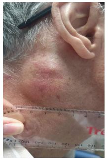
Localized cervical lymphadenopathy is a rare manifestation of diffuse cutaneous Systemic Sclerosis (dcSSc), and its diagnosis can be challenging given the wide range of diseases associated with this complaint. Clinical, laboratory, imaging and histological assesment are fundamental for accurate clinical reasoning. A 62-year-old man, former smoker, with a history of atrial fibrillation under anticoagulation, arterial hypertension and diffuse cutaneous systemic sclerosis with multi-organ involvement, with Autoimmune Diseases consultations regularly. The patient reported a mass in the right retroauricular region that had been present for approximately three weeks, without an apparent cause, and an involuntary weight loss of 2 kgs over one month, denying other symptoms. On physical examination, a painful, stony-hard swelling was palpated, adherent to the deep planes, with a diameter of 2.5 cm. Given the clinical context, analytical and imaging studies were performed, revealing no new alterations. The ultrasound study showed “the presence of some lymphadenopathies at the right mandibular angle, the largest measuring 37 mms, with thickening of the sternocleidomastoid muscle”. The patient was seen in the Maxillofacial Surgery consultation two months later for a biopsy, but by that time, no mass was observed. The patient was discharged, and lymphadenopathy was assumed to be related in the context of diffuse systemic sclerosis. The appearance of localized lymphadenopathy in the context of diffuse cutaneous systemic sclerosis is a rare finding, often underdiagnosed due to the low specificity of this complaint. Although histological evaluation is essential, laboratory and imaging studies allow the exclusion of most differential diagnoses. The therapeutic approach depends on the disease`s activity and the patient`s overall condition. The case reinforces the importance of recognizing alarm signs in the evaluation of lymphadenopathy and highlights the relevance of addressing the patient and their complaints individually and within their clinical context to achieve an accurate diagnostic and therapeutic approach.