
Journal of Clinical Images and Medical Case Reports
ISSN 2766-7820
Clinical Image - Open Access, Volume 6
An image of the xanthogranuloma of the plexus choroid
Nada Adjou*; Majda Ankri; Soukaina Jabour; Amal Lahfidi; Firdaous Touarssa; Meryem Fikri; N El Kettani; Mohamed Jidanne
Neuroradiology Department of the Specialty Hospital of Rabat, Mohamed V University Rabat, Morocco.
*Corresponding Author : Nada Adjou
Neuroradiology Department of the Specialty
Hospital of Rabat, Mohamed V University Rabat,
Morocco.
Email: docnada736@gmail.com
Received : Feb 25, 2025
Accepted : Mar 19, 2025
Published : Mar 26, 2025
Archived : www.jcimcr.org
Copyright : © Adjou N (2025).
Keywords: Xanthogranuloma; Plexus choroid; Cyst.
Citation: Adjou N, Ankri M, Jabour S, Lahfidi A, Touarssa F, et al. An image of the xanthogranuloma of the plexus choroid. J Clin Images Med Case Rep. 2025; 6(3): 3525.
Description
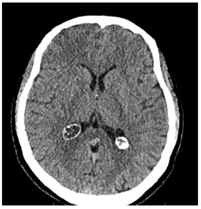
A 34 year old women consulted in emergency in our department for intense headache with vomiting, and bilateral papilledema in the ophtalmologic examination, in front of these symptoms a brain CT-scan was performed. We observed a well defined oval formation of fluid density in the occipital horn of the right lateral ventricle with calcified wall in favor of a xanthogranuloma of the choroid plexus (Figure 1). The first description of an xanthogranuloma was reported by Blunner in 1900 as cholesteatomatous endothelioma [1], since then they have been reported incidentally mostly in the lateral ventricules. Xanthogranuloma of the choroid plexus can cause obstructive hydrocephalus, and patients may develop neurological symptoms such as a headache, papilledema, gait instability, cognitive impairment, and urinary disturbances [1]. XGRs represent tissue reactions with pseudotumor aspect and are composed of cholesterol clefts and inflammatory cells such as foamy cells, histiocytes, multinucleated foreign body giant cells, hemosiderin deposits and fibrous proliferation [2]. CT findings of choroid plexus xanthogranulomas in the litterature are variable from hypo, iso and hyperdense, representing the heterogeneity in the content of these lesions. They are round and smooth-walled [1]. There etiology remains controversial, some authors relate the condition to an inflammatory response to the proliferation and invagination of the choroid plexus and neuroepithelial cyst transformation, and other reports suggest a developmental origin or high levels of lipids in the blood [1,3,4].
References
- Miranda P, Lobato RD, Ricoy JR, Lagares A, Ramos A. Xanthogranuloma of the choroid plexus of the third ventricle : Case report and littérature review, Neurocirugia (Astur). 2005; 6: 518- 22.
- Gontier MF. Pathological anatomy of tumors of the third ventricule. Neurochirurgie. 2000; 46: 257-67.
- Gaskill SJ, Saldivar V Rutman J, Marlin AE. Giant bilateral xanthogranulomas in a child: Case report.Neurosurgery. 1992; 31: 114-7.
- Bruck W sander U, Blanckenberg P, Friede RL. Symptomatic xanthogranuloma of choroid plexus with unilateral hydrocephalus. Case report . J Neurosurg. 1991; 5: 324.