Journal of Clinical Images and Medical Case Reports
ISSN 2766-7820
Clinical Image - Open Access, Volume 6
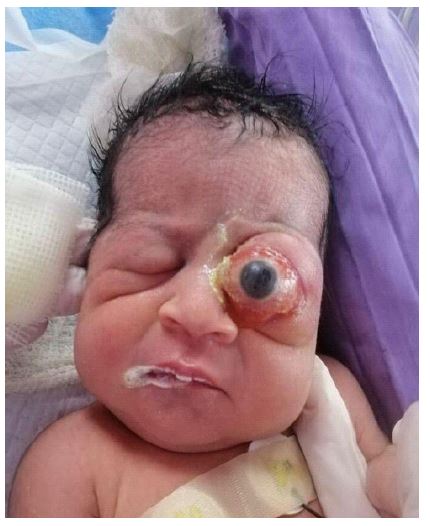
Congenital mature orbital teratoma in a neonate
*Corresponding Author : Mohamad Hammoud
Department of Pediatrics, American University of Beirut Medical Center Street, Hamra- Beirut, Lebanon.
Tel: +961-7-079-7154; Email: mh350@aub.edu.lb
Received : Mar 06, 2025
Accepted : Mar 26, 2025
Published : Apr 02, 2025
Archived : www.jcimcr.org
Copyright : © Hammoud M (2025).
Citation: Hammoud M. Congenital mature orbital teratoma in a neonate. J Clin Images Med Case Rep. 2025; 6(4): 3537.
Description
A 1-day-old boy, born at 39 weeks gestational age without relevant prenatal history, presented with a large vascularized pyroptotic mass occupying the left midface. No family history of congenital deformities, no parents’ consanguinity and no teratogenic drugs used during pregnancy. Shortly after birth, Ultrasound and computed tomography showed a heterogeneous mass mixed with peripheral calcific changes, occupying the retro-globar space of the eye, particularly the intra-Conal orbital compartment. The ocular globe is shifted anteriorly resulting in considerable exophthalmos associated with thickening of the respective eyelid without extension to surrounding structures. No brain deformities are seen. Magnetic resonance of the orbit showed a large mixed cystic and solid left retro-orbital mass filling the intraconal space and exerting mass effect on the extra- ocular muscles and displacing the left optic nerve as well. Clinical and imaging features were consistent with congenital teratoma of the orbit. The patient underwent an urgent orbitotomy with excision of the retro-orbital lesion without any enucleation or exenteration. The tumor was resected, eye globe was restored inside the orbit and the optic nerve was spared with no intraoperative complications. Frozen section pathology and histopathological section confirmed the diagnosis of mature orbital teratoma. Postoperatively he was given antibiotics and analgesics. The patient was discharged on the 5th postoperative day without any complications.