
Journal of Clinical Images and Medical Case Reports
ISSN 2766-7820
Case Report - Open Access, Volume 6
Radiation-induced morphea occurring outside the irradiated field in a patient with squamous cell carcinoma of the anus: A case report
Eric D Young1; Andrew C Hoover2*
1Departments of Cancer Biology, University of Kansas Medical Center, Kansas City, KS 66160, USA.
2Radiation Oncology, University of Kansas Medical Center, Kansas City, KS 66160, USA.
*Corresponding Author : Andrew C Hoover
Radiation Oncology, University of Kansas Medical Center, Kansas City, KS 66160, USA.
Email: ahoover2@kumc.edu
Received : Mar 10, 2025
Accepted : Apr 02, 2025
Published : Apr 09, 2025
Archived : www.jcimcr.org
Copyright : © Hoover AC (2025).
Abstract
Background: Radiation-Induced Morphea (RIM) is a rare sequela of radiation therapy occurring most frequently in patients with breast cancer and is usually identified within the irradiated field. Diagnosis of RIM is challenging and frequently a diagnosis of exclusion. Patients who have received radiation therapy may present with a rash from a number of similar-appearing conditions including cancer recurrence, new carcinoma, cellulitis, lymphedema and chronic radiation dermatitis or recall dermatitis. Much remains to be understood about the pathophysiology of RIM. Currently, the natural history is thought to involve an inflammatory phase causing rash and an atrophic phase terminating in fibrosis. To date, very few verified cases of RIM have been reported in patients with cancers other than breast cancer and examples of RIM occurring outside the irradiated field are rare. Here, we report a case of RIM occurring outside the irradiated field in a patient with Squamous Cell Carcinoma (SCC) of the anus.
Case presentation: A 52-year-old female with a history of poorly differentiated basaloid SCC of the anus presented with a rash approximately 29 months after radiation therapy. The patient had been treated for her cancer with external beam radiation (EBRT, 54 Gray in 30 fractions) and concurrent chemotherapy. A multifocal, erythematous rash progressed to stiffened plaques in on her legs, abdomen, face and pelvis. Further dermatologic workup revealed a diagnosis of RIM. The patient initially underwent 2-month course of Clindamycin-Tretinoin treatment and topical steroids with limited results. This was followed by treatment with Dapsone and Prednisone, which unfortunately produced minimal improvement of sclerotic plaques.
Conclusion: While RIM is most frequently associated with patients who have a history of breast cancer, it also occurs in patients with other types of cancer. Differential diagnosis of a rash in patients with a history of radiation therapy is broad and often challenging. Therefore, diagnosis of RIM in patients with cancers occurring outside the breast may be underreported. RIM should be recognized as a rare but important consideration in the differential diagnosis of patients treated with radiotherapy and can occur outside the irradiated field in patients with a history of SCC of the anus.
Keywords: Morphea; Radiation-induced morphea; Anal cancer; Squamous cell carcinoma; Radiation induced morphea; Localized scleroderma.
Abbreviations: CT: Computerized Tomography; EBT: External Beam Radiation Therapy; Gy: Gray; IHC: Immunohistochemistry; MRI: Magnetic Resonance Imaging; PET: Positron-Emission Tomography; PIM: Post-Irradiation Morphea (Synonymous With RIM); RIM: Radiation-Induced Morphea; SCC: Squamous Cell Carcinoma.
Citation: Young ED, Hoover AC. Radiation-induced morphea occurring outside the irradiated field in a patient with squamous cell carcinoma of the anus: A case report. J Clin Images Med Case Rep. 2025; 6(4): 3545.
Introduction
Radiation-Induced Morphea (RIM) is a rare sequela of radiation therapy, which to date has been most frequently described in patients with a history of breast cancer [1,2]. The association of morphea with radiation treatment was elucidated by Colver et al. in 1989 in which the majority of patients had a history of breast cancer, but also described patients with cancer of the head and neck and endometrial adenocarcinoma [3,4]. Since 1989, RIM has also been reported in a small number of patients with other cancer types including cancer of the head and neck [5,6], cervix [5], endometrium [7,8], and lymphoma [9]. While RIM most frequently occurs within the irradiated field [1,10], there are examples of it occurring outside the irradiated field [4,7,8,10-12]. RIM is an idiopathic inflammatory condition of the skin. The pathogenesis is currently thought to involve an initial inflammatory phase resulting in increased fibroblast activation and consequent fibrosis which is then followed by a resolving and atrophic phase that can cause pain, contracture and changes in skin coloration (hypopigmentation or hyperpigmentation) of the affected area [13]. Current treatment options for RIM include immunosuppressants, anti-fibrotics, immune stimulants and photopheresis, which are of variable reported efficacy in resolving the signs and symptoms of RIM [14]. Differential diagnosis of a rash in a patient after receiving radiation therapy is broad and identifying the etiology can be challenging [15]. Appreciating that RIM can occur in patients with many types of cancer can contribute to accurate diagnosis of future patients. Here, we report what we believe to be the first report of RIM occurring outside the irradiated field in a patient treated for SCC of the anus.
Case presentation
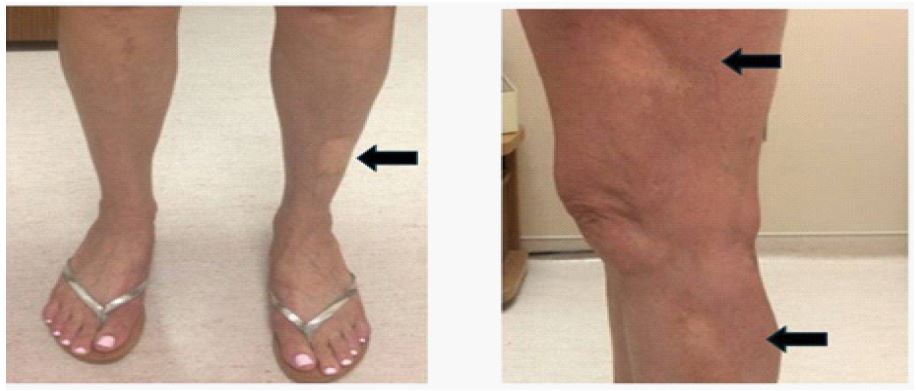
Here, we describe a case of RIM occurring in a patient with a history of SCC of the anus without any history of breast cancer. The patient was a 52-year-old Caucasian female diagnosed with SCC of the anus (stage IIIB; T4, N1, M0) after workup for rectal bleeding and dyschezia. Her Body Mass Index (BMI) was within normal range. Colonoscopy revealed a 3-4 cm palpable rectal mass. Magnetic Resonance Imaging (MRI) and Positron-Emission Tomography (PET) imaging showed invasion of rectum and posterior vaginal wall with enlarged and hypermetabolic rectal and presacral lymph nodes without evidence of distant metastatic disease. Excisional biopsy revealed invasive moderate to poorly differentiated basaloid SCC that was positive for p40 and p63 by Immunohistochemistry (IHC). After biopsy, patient was treated definitively with External Beam Radiation (EBRT) (54 Gray in 30 fractions) with concurrent chemotherapy (mitomycin and fluorouracil). Monitoring was achieved by post-therapy PET and Computerized Tomography (CT) imaging in addition to serial anoscopies and exams under anesthesia. Subsequent biopsies were negative for malignancy. The patient presented to her dermatologist with “red spots” on her skin 29 months after radiation therapy. These rashes progressed to sclerotic and hypopigmented plaques on her left leg and thigh, abdomen, face and pelvis. Rashes were located outside the previously irradiated field (Figure 1). Patient did have a history of Hashimoto’s thyroiditis and subsequent hypothyroidism, but denied any other history of vasculitis, autoimmune or inflammatory conditions. Other medical history included hypertension, cervical disk herniation, renal and hepatic cysts, anxiety and irritable bowel syndrome. The patient had previously undergone tonsillectomy as a child. Patient was previously treated for cervical disk herniation, Cesarean section, removal of a sessile colon polyp, and removal of an ovarian cyst. Patient was a non-smoker, consumed 5-7 alcoholic drinks per week and used no other drugs. Family history was positive for depression, cardiovascular disease and thyroid cancer. For her skin lesions, patient was treated with a 2-month course of Clindamycin-Tretinoin and topical steroids with limited results. This was followed by treatment with Dapsone and Prednisone, which unfortunately provided minimal improvement of sclerotic plaques.
Discussion and conclusion
Here, we present what we believe to be the first report of RIM in a patient treated for SCC of the anus and one of few reported cases of RIM occurring outside the irradiated field. It has been observed that cases of RIM presenting outside the irradiated field tend to occur in the lower extremities [4,7,8,10], and our patient’s presentation is consistent with this pattern. The manifestation of RIM after radiotherapy in a region outside the irradiated field suggests that RIM may be a systemic response to an inciting inflammatory stimulus, such as radiation. But why the lower extremities might be predisposed to developing RIM lesions is not known. The fact that morphea also occurs, albeit less frequently, as a spontaneous condition in the absence of radiotherapy [16] might suggest that there is a genetic contribution to RIM which could predispose an individual to this exaggerated inflammatory response. Numerous disease associations have been suggested that may predispose an individual to developing RIM, such as breast implantation, autoimmune disease, smoking and obesity [15]. Indeed, our patient did report a history of autoimmune disease (Hashimoto’s thyroiditis), which may have been a contributing factor in her developing RIM. However, the development of RIM with a single risk factor demonstrates that the disease remains stochastic with respect to which patients are affected and that the biological mechanism of this disease is at present poorly understood. While RIM is reported frequently in patients with a history of breast cancer, it is not known if cancer type is an independent risk factor for developing RIM [2]. Breast cancer’s association with many of the aforementioned potential risk factors might confound their interpretation. Other explanations for the frequent association of RIM in patients with breast cancer might be the relatively high prevalence of breast cancer as opposed to other cancer types and frequent use of external beam radiation in these patients with breast cancer, a heightened bodily awareness in patients treated for breast cancer, or attention bias toward diagnosis of the condition in patients with a history of breast cancer as compared to patients with other cancer types. The resolution of these important questions depends upon continued reporting of patients with RIM. While it is a rare disease, its potential for physical and psychological morbidity due to scarring and contracture is substantial and warrants further study.
Declarations
Publication ethics: Publication of de-identified patient data was approved by the University of Kansas Medical Center ethics committee.
Conflicts of interest: The authors declare that they have no competing financial interests.
References
- NR Bleasel, KM Stapleton, C Commens, VA Ahern. Radiation-induced localized scleroderma in breast cancer patients, (in eng), Australas J Dermatol. 1999; 40(2): 99-102. doi: 10.1046/j.1440-0960.1999.00330.x.
- A Alhathlool, R Hein, C Andres, J Ring, B Eberlein. Post-Irradiation Morphea: Case report and review of the literature, (in eng), J Dermatol Case Rep. 2012; 6(3): 73-7. doi: 10.3315/jdcr.2012.1106.
- GB Colver, A Rodger, PS Mortimer, JA Savin, SM Neill, et al. Post-irradiation morphoea, (in eng), Br J Dermatol. 1989; 120(6): 831-5. doi: 10.1111/j.1365-2133.1989.tb01382.x.
- MR Ardern-Jones, MM Black. Widespread morphoea following radiotherapy for carcinoma of the breast. (in eng), Clin Exp Dermatol. 28(2): 160-2. Mar 2003, doi: 10.1046/j.1365-2230.2003.01186.x.
- M Abu-Shakra, F Guillemin, P Lee. Cancer in systemic sclerosis, (in eng), Arthritis Rheum. 1993; 36(4): 460-4. doi: 10.1002/art.1780360405.
- SG Cooper, JW Denham. Progressive systemic sclerosis (diffuse scleroderma) and radiotherapy, (in eng), Br J Radiol. 1990; 63l(754): 804-5. doi: 10.1259/0007-1285-63-754-804.
- H Ullén, E Björkholm. Localized scleroderma in a woman irradiated at two sites for endometrial and breast carcinoma: A case history and a review of the literature, (in eng), Int J Gynecol Cancer. 2003; 13(1): 77-82. doi: 10.1046/j.1525-1438.2003.13006.x.
- BN Akay, H Sanli, AO Heper. Postirradiation linear morphoea, (in eng), Clin Exp Dermatol. 2010; 35(4): 106-8. doi: 10.1111/j.1365-2230.2009.03717.x.
- KJ Smith, J Yeager, HG Skelton. Localized scleroderma in breast cancer patients treated with supervoltage external beam radiation: Radiation port scleroderma, (in eng), J Am Acad Dermatol. 1997; 37(5-1): 806-8. doi: 10.1016/s0190-9622(97)70130-0.
- J Kushi, ME Csuka. Generalized morphea after breast cancer radiation therapy, (in eng), Case Rep Rheumatol. 2011; 951948. doi: 10.1155/2011/951948.
- S Balegar, DK Mishra, S Chatterjee, S Kumari, AK Tiwary. Generalized Morphea following Radiotherapy for an Intracranial Tumor, (in eng), Indian J Dermatol. 2016; 61(5): 581. doi: 10.4103/0019-5154.190132.
- K Yanaba, Y Umezawa, H Nakagawa. A case of radiation-induced generalized morphea with prominent mucin deposition and tenderness, (in eng), Am J Case Rep. 2015; 16: 279-82. doi: 10.12659/ajcr.893481.
- I Badea, M Taylor, A Rosenberg, M Foldvari. Pathogenesis and therapeutic approaches for improved topical treatment in localized scleroderma and systemic sclerosis, (in eng), Rheumatology (Oxford). 2009; 48(3): 13-21. doi: 10.1093/rheumatology/ken405.
- M Spalek, J Jonska-Gmyrek, J Gałecki. Radiation-induced morphea-a literature review, (in eng), J Eur Acad Dermatol Venereol. 2015; 29(2): 197-202. doi: 10.1111/jdv.12704.
- A Mittal, V Mittal, G Panse, JN Choi, BY Kwong, et al. Radiation-induced morphea: Association with autoimmune comorbidities, severity, and response to therapy, (in eng), J Am Acad Dermatol. 2019; 81(1): 260-262. doi: 10.1016/j.jaad.2019.02.039.
- LS Peterson, AM Nelson, WP Su, T Mason, WM O’Fallon, et al. The epidemiology of morphea (localized scleroderma) in Olmsted County 1960-1993, (in eng), J Rheumatol. 1997; 24(1): 73-80.