Journal of Clinical Images and Medical Case Reports
ISSN 2766-7820
Clinical Image - Open Access, Volume 6
Non-ulcerated gastrointestinal stromal tumor presenting with hematemesis
Chaima Jioua1*; Salma Azammam2; Jihane Benass2; Reda Berraida2; Imane Moulim1; Sanaa Berrag1; Fouad Nejjari1; Tarik Adioui1; Mouna Tamzaourte1
1Department of Gastroenterology I, Mohamed V Military Training Hospital, Rabat, Morocco.
2Department of Gastroenterology II, Mohamed V Military Training Hospital, Rabat, Morocco.
*Corresponding Author : Chaima Jioua
Department of Gastroenterology I, Mohamed V Military Training Hospital, Rabat, Morocco.
Email: chaimaej.cj@gmail.com
Received : Mar 10, 2025
Accepted : Apr 02, 2025
Published : Apr 09, 2025
Archived : www.jcimcr.org
Copyright : © Jioua C (2025).
Citation: Jioua C, Azammam S, Benass J, Berraida R, Moulim I, et al. Non-ulcerated gastrointestinal stromal tumor presenting with hematemesis. J Clin Images Med Case Rep. 2025; 6(4): 3546.
Description
A 39 year-old man with no significant medical history presented to the emergency department following three episodes of hematemesis. He reported several weeks of intermittent epigastric pain and fatigue but denied any weight loss. There was no history of Non Steroidal Anti-Inflammatory Drug (NSAID) use, anticoagulation, or alcohol consumption. On examination,he was pale, tachycardic (112 bpm), and hypotensive (90/60 mmHg), Physical examination demonstrated epigastric tenderness without palpable masses or peritoneal signs. Laboratory investigations revealed anemia (Hb: 8 g/dL), coagulation profiles and liver function tests were within normal limits. The patient received rapid crystalloid infusion and transfusion of two units of packed red blood cells, and he received a bolus of Proton Pump Inhibitor (PPI), followed by continuous intravenous infusion.
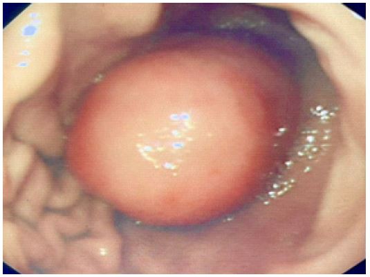
Urgent esophagogastroduodenoscopy revealed a submucosal, protruding lesion in the gastric fundus without a visible ulceration, or active bleeding. No hemostatic intervention was performed due to the absence of an active bleeding point.
The evolution has been marked by recurrent melena and a drop in hemoglobin to 7 g/dL over the next 48 hours. Given ongoing bleeding, surgical intervention was planned. The patient underwent laparoscopic resection of the tumor. Intraoperatively, the mass was well-circumscribed, vascular, and covered with intact mucosa, with evidence of intratumoral hemorrhage but no external ulceration. The mass was successfully resected with negative margins. Histopathology confirmed Gastrointestinal Stromal Tumor (GIST) with CD117 and DOG1 positivity. Given the intermediate-risk classification, adjuvant imatinib (400 mg/day) was initiated. At 6-month follow-up, the patient remained asymptomatic with no signs of recurrence.