
Journal of Clinical Images and Medical Case Reports
ISSN 2766-7820
Clinical Image - Open Access, Volume 6
A case of pseudoepitheliomatous hyperplasia mimicking squamous cell carcinoma over gluteal region: Clinical image
Sushendra T*; Hetal Nakrani2; Ponnuleksmi3; Vishnupriya3
1Professor, Department of Shalya Tantra, JS Ayurveda Mahavidyalaya, Nadiad, Gujarat, India.
2Reader, Department of Shalya Tantra, JS Ayurveda Mahavidyalaya, Nadiad, Gujarat, India.
3Lecturer, Department of Shalya Tantra, JS Ayurveda Mahavidyalaya, Nadiad, Gujarat, India.
*Corresponding Author : T Sushendra
Professor, Department of Shalya Tantra, JS Ayurveda Mahavidyalaya, Nadiad-387001, Gujarat, India.
Tel: 9900907644;
Email: sushendra84@gmail.com
Received : Mar 21, 2025
Accepted : Apr 09, 2025
Published : Apr 16, 2025
Archived : www.jcimcr.org
Copyright : © Sushendra T (2025).
Citation: Sushendra T, Nakrani H, Ponnuleksmi, Vishnupriya. A case of pseudoepitheliomatous hyperplasia mimicking squamous cell carcinoma over gluteal region: Clinical image. J Clin Images Med Case Rep. 2025; 6(4): 3555.
Description
Pseudoepitheliomatous hyperplasia (PEH) is a benign condition characterized by the proliferation of the epidermal and adnexal epithelium, closely resembling Squamous Cell Carcinoma (SCC). PEH is often confused with SCC [1]. We describe a case upon clinical suspicion of squamous cell carcinoma and a biopsy was conducted, confirming the diagnosis of PEH, which was then managed accordingly.
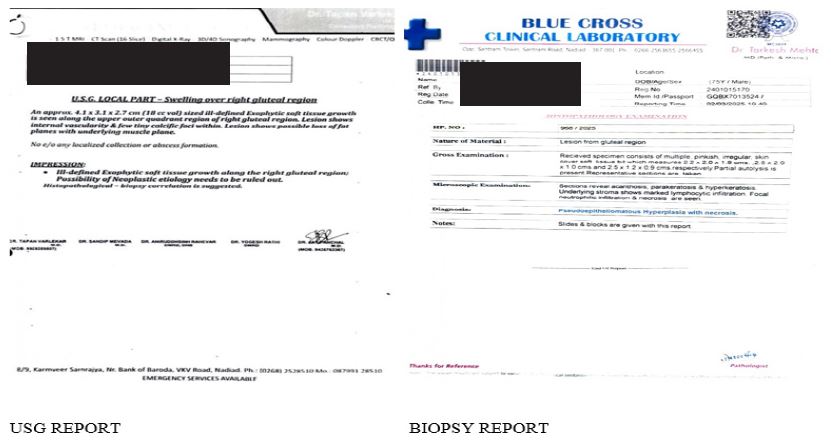
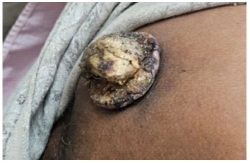
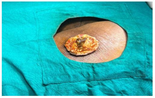
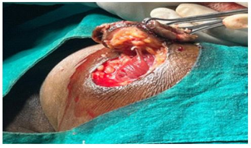
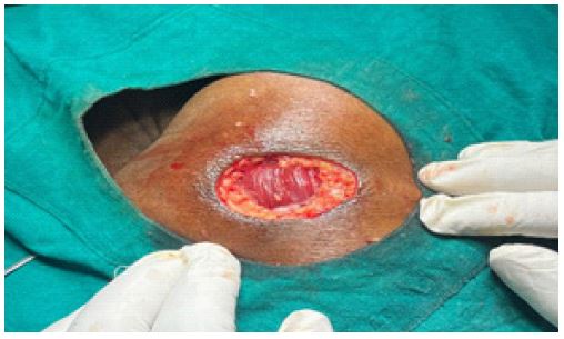
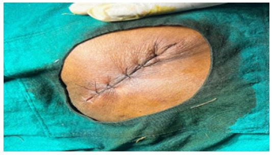
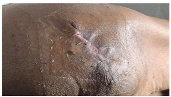
A 60 yrs old male patient non diabetic, non-hypertensive came to OPD presenting with swelling over right gluteal region associated with pain for 2 months. Initially it appeared as small as boil like and later rapidly increased in size. Patient had pain and discomfort while sitting for long time. There was no history of similar complaints in the past or in the family. On examination the mass measured about 4 cm x 3 cm x 3 cm with everted margins, with ulceration, necrotic tissue and fumigation over the center. On palpation slight tenderness with mild induration surrounding the swelling. Regional lymph nodes were normal on palpation. Initially suggested USG of the local part which showed ill-defined exophytic soft tissue growth along right gluteal region. In order to rule out malignancy, biopsy was done which reported as Pseudoepitheliomatous hyperplasia. After the confirmation of the diagnosis the mass was excised under local anesthesia and was closed with Ethilon 3-0 with mattress suture and the suture was removed after 8 days and wound healed complete after 15 days.
References
- Chakrabarti S, Chakrabarti PR, Agrawal D, Somanath S. Psuedoepitheliomatous hyperplasia: a clinical entity mistaken for squamous cell carcinoma. J Cotan Aesthete Surg. 2014; 7(4): 232-4. doi: 10.4103/0974-2077.150787.