Journal of Clinical Images and Medical Case Reports
ISSN 2766-7820
Clinical Image - Open Access, Volume 6
Pulmonary dirofilariasis on trans-thoracic needle biopsy
*Corresponding Author : Vivek Sinanan
Baylor College of Medicine, Baylor Plaza, Houston, TX 77030 USA.
Tel: 9083271728;
Email: vivek.sinanan@bcm.edu
Received : Mar 25, 2025
Accepted : Apr 14, 2025
Published : Apr 21, 2025
Archived : www.jcimcr.org
Copyright : © Sinanan V (2025).
Citation: Sinanan V. Pulmonary dirofilariasis on trans-thoracic needle biopsy. J Clin Images Med Case Rep. 2025; 6(4): 3562.
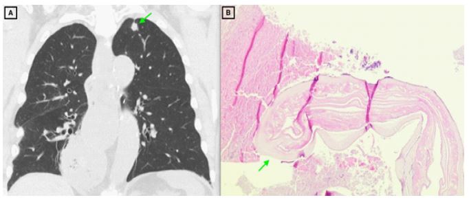
Description
A 79 year-old male was incidentally found to have a 14 mm spiculated nodule in the left upper lobe on CT imaging of the cervical spine (Figure 1A). He had no respiratory symptoms and is a never smoker. PET-CT demonstrated mild FDG uptake and subsequent CT-guided trans-thoracic needle biopsy only showed extensive necrosis. A repeat biopsy showed atypical cells, and necrotic fragments of dirofilarial were identified during correlation with tissue from the first biopsy (Figure 1B).
Dirofilarial are vector-borne nematodes that cause dirofilariasis. Infected mosquitos transmit the larvae to the definitive host, dogs, where they infect the right ventricle and pulmonary arteries. Humans are poor hosts, resulting in larvae death and embolization to the lungs where they can present as pulmonary nodules or infarcts. Most cases are diagnosed via surgical biopsy due to the concerning appearance of the associated nodules. Our patient represents an unusual diagnosis made via trans-thoracic needle biopsy.