
Journal of Clinical Images and Medical Case Reports
ISSN 2766-7820
Clinical Image - Open Access, Volume 6
Behcet’s disease misdiagnosed as a soft tissue infection
Mert Tokatli1*; Oguz Abdullah Uyaroglu2
1Department of Internal Medicine, Faculty of Medicine, Hacettepe University, Ankara, Turkey.
2Department of General Internal Medicine, Faculty of Medicine, Hacettepe University, Ankara, Turkey.
*Corresponding Author : Mert Tokatli
Department of Internal Medicine, Faculty of Medicine, Hacettepe University, 06100, Altindag/Ankara, Turkey.
Tel: +90 544 205 5400;
Email: merttokatli17@gmail.com
Received : Mar 28, 2025
Accepted : Apr 15, 2025
Published : Apr 22, 2025
Archived : www.jcimcr.org
Copyright : © Tokatli M (2025).
Citation: Tokatli M, Uyaroglu OA. Behcet’s disease misdiagnosed as a soft tissue infection. J Clin Images Med Case Rep. 2025; 6(4): 3563.
Description
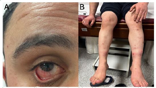
A 27-year-old male patient was admitted to our clinic, presenting with complaints of chronic leg sores that had persisted for one and a half years. The wounds did not heal despite various antibiotic treatments. Additionally, the patient had a history of enoxaparin use due to thrombosis in the lower extremity of the right leg. The patient was observed to have redness in the right eye (Figure 1A) and a history of uveitis. Painful, erythematous, warm, nodular lesions on the anterior aspect of bilateral legs were evaluated as erythema nodosum (Figure 1B). Diffuse papulopustular lesions all over the body, aphthous plaque on the lower lip, and genital papulopustular lesions were present. Punch biopsy from the leg was reported as panniculitis (lobular predominance) and small vessel vasculitis, and the Pathergy test was positive. Behçet’s Disease, also known as Behçet’s Syndrome, is a systemic vasculitis, an inflammation of the blood vessels. Recurrent mouth ulcers, genital ulcers, and skin lesions characterize this chronic autoimmune disease. It is one of the rare diseases that can affect both small and large blood vessels. The condition was first described in 1937 by Hulusi Behçet [1]. The International Working Group criteria for Behçet’s disease, in addition to recurrent oral ulcers, the presence of two of the following is diagnostic: eye involvement, skin lesions, and a positive pathergy test [2]. Oligoarticular arthritis, superficial and deep vein thrombosis, central nervous system involvement, and gastrointestinal involvement may also be seen. HLA-B51 positivity is found in 60-70% of patients. The most serious complication is vision loss. Organ involvement is crucial in treatment. Topical steroids, thalidomide, and apremilast are effective for mucocutaneous lesions, while colchicine can be added if arthritis is present. Systemic steroids, azathioprine, cyclosporine, and anti-TNF therapies are utilized when uveitis is present [3]. Cyclophosphamide may be administered in cases of pulmonary artery vasculitis. Behçet’s disease should be considered when eye involvement and persistent skin lesions are observed.
Declarations
Patient consent: Consent was obtained from the patient to publication of their information and images.
Competing interest: None declared.
Funding: No funding.
References
- Behcet H. Uber rezidiverende aphthose durch ein virus verursachte Geschwure am Mund, am Auge und an den Genitalien. 1937.
- Davatchi F, et al., The International Criteria for Behçet’s Disease (ICBD): A collaborative study of 27 countries on the sensitivity and specificity of the new criteria. Journal of the European Academy of Dermatology and Venereology, 2014; 28(3): 338-347.
- Karadag O, EC Bolek. Management of Behcet’s syndrome. Rheumatology, 2020. 59(3): 108-117.