
Journal of Clinical Images and Medical Case Reports
ISSN 2766-7820
Short Report - Open Access, Volume 6
Proteus mirabilis as a cause of intertrigo: The value of UV dermoscopy
Meryem Soughi1*; Aimane Zaimi2; Zakia Douhi1; Sara Elloudi1; Hanane Baybay1; Fatima Zahra Mernissi1
1Dermatology Department, University Hospital Hassan II, URL CNRST N15; Human Pathology, Biomedicine and Environment Laboratory; Faculty of Medicine, Pharmacy and Dental Medicine of Fez. Sidi Mohamed Ben Abdellah University, Fez, Morocco.
2Dermatology Department, Faculty of Medicine, Pharmacy and Dental Medicine, University Hospital Hassan II Fez, Morocco.
*Corresponding Author : Meryem Soughi
Faculty of Medicine, Pharmacy and Dental Medicine of Fez. Sidi Mohamed Ben Abdellah University, Fez, Morocco.
Tel: 00212611936739;
Email: msoughi@gmail.com
Received : Mar 21, 2025
Accepted : Apr 18, 2025
Published : Apr 25, 2025
Archived : www.jcimcr.org
Copyright : © Soughi M (2025).
Abstract
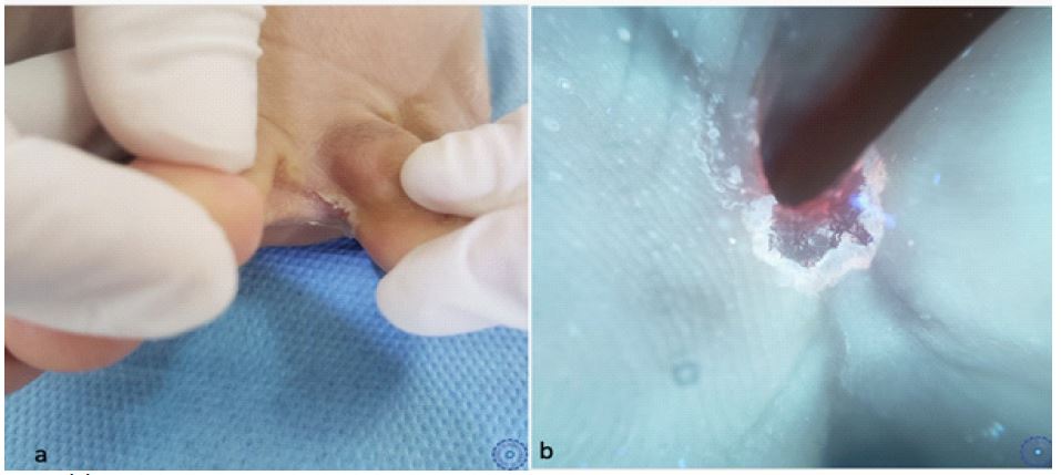
A 50-year-old patient with no significant medical history, presented with an intertrigo resistant to antifungal treatment. Dermatological examination revealed a dry intertrigo with fine, powdery scales and a peripheral collarette in the fourth interdigital space, Under Ultraviolet-induced fluorescence (UVF), a coral-red fluorescence was observed in the fold, with more pronounced scales and a collarette, Bacteriological and mycological examination of the scales identified a Gram-negative bacillus, spores, and mycelial filaments. Culture results confirmed the presence of Proteus mirabilis and Candida albicans. The patient was treated with a topical antifungal and antibiotic, leading to significant improvement. This case highlights the value of UV dermoscopy in entomodermoscopy and its role in diagnostic orientation. It also underscores the presence of an unusual pathogen, Proteus mirabilis, alongside Candida as a causative agent of intertrigo.
Keywords: Dermoscopy; UV dermoscopy; Proteus mirabelis; Candida albicans; Intertrigo.
Citation: Soughi M, Zaimi A, Douhi Z, Elloudi S, Baybay H, et al. Proteus mirabilis as a cause of intertrigo: The value of UV dermoscopy. J Clin Images Med Case Rep. 2025; 6(4): 3569.
Background
UVF dermoscopy is an innovative dermoscopic technique that utilizes a UV light source at 365 nm to induce fluorescence in skin chromophores [1]. We report a case of interdigital intertrigo caused by Proteus mirabilis, in which the diagnosis was guided by UVF dermoscopy.
Case presentation
A 50-year-old patient with no significant medical history, presented with an intertrigo resistant to antifungal treatment. Dermatological examination revealed a dry intertrigo with fine, powdery scales and a peripheral collarette in the fourth interdigital space (Figure 1a), Under UVF dermoscopy, a coral-red fluorescence was observed in the fold, with more pronounced scales and a collarette (Figure 1b), Bacteriological and mycological examination of the scales identified a Gram-negative bacillus, spores, and mycelial filaments. Culture results confirmed the presence of Proteus mirabilis and Candida albicans. The patient was treated with a topical antifungal and antibiotic, leading to significant improvement.
Discussion
Cutaneous infections caused by Proteus mirabilis can present in various clinical forms, ranging from acute cellulitis, to macular, papular, or erythematous lesions. These manifestations can sometimes mimic Pseudomonas aeruginosa, making clinical diagnosis challenging [2]. In this context, UVF dermoscopy are innovative diagnostic tools capable of identifying clues that are not discernible with conventional dermoscopy [2,3]. UVF dermoscopy is based on the Stokes shift phenomenon corresponding to the emission of visible fluorescent photons by chromophores excited by UV light [2,3]. In practical applications, UVF dermoscopy does not reveal any fluorescent signals in Candida-induced intertrigo. However, a green fluorescence is observed in Pseudomonas aeruginosa infections due to the secretion of pyoverdine, while a red fluorescence is typically associated with Corynebacterium infections, attributed to the production of coproporphyrin III [2,3].
In our case, a red fluorescence was observed, suggesting a bacterial origin. However, bacteriological analysis confirmed the presence of Proteus mirabilis, a bacterium known for its production of hydrogen sulfide (H₂S), but not reported associated with red fluorescence. The unexpected fluorescence led us to perform an additional microbiological assessment, ultimately guiding a therapeutic adjustment with the introduction of targeted antibiotic treatment. Although the exact mechanism underlying the observed red fluorescence remains unclear, our findings highlight the potential role of UVF dermoscopy as a valuable diagnostic aid in cutaneous infections.
Conclusion
The purpose of presenting this case is to highlight the value of UVF dermoscopy in entomodermoscopy and its role in diagnostic orientation. It also underscores the presence of an unusual pathogen, Proteus mirabilis, alongside Candida as a causative agent of intertrigo.
References
- Errichetti E, Pietkiewicz P, Bhat YJ, Salwowska N, Szlązak P, Stinco G. Diagnostic accuracy of ultraviolet-induced fluorescence dermoscopy in non-neoplastic dermatoses (general dermatology): A multicentric retrospective comparative study. J Eur Acad Dermatol Venereol. 2025; 39(1): 97-108. doi: 10.1111/jdv.19795. Epub 2024 Jan 30.
- Krajden S, Deitel M, Fuksa M. Blackened toes caused by Proteus mirabilis infection. Can Med Assoc J. 1982; 127(9): 869-70.
- Pietkiewicz P, Navarrete-Dechent C, Togawa Y, Szlązak P, Salwowska N, Marghoob AA, et al. Applications of Ultraviolet and Sub-ultraviolet Dermatoscopy in Neoplastic and Non-neoplastic Dermatoses: A Systematic Review. Dermatol Ther (Heidelb). 2024; 14(2): 361-390. doi: 10.1007/s13555-024-01104-4. Epub 2024 Feb 15.