
Journal of Clinical Images and Medical Case Reports
ISSN 2766-7820
Short Report - Open Access, Volume 6
Short report of an intramuscular hibernoma as an incidental finding in 18F-FDG PET/CT
Jesús López Urdaneta1; Henrik Fagman2; Sonja Kjartansdóttir3; Pablo Borrelli1*
1Nuclear Medicine Session, Sahlgrenska University Hospital, Gothenburg, Sweden.
2Department of Clinical Pathology, Sahlgrenska University Hospital, Gothenburg, Sweden.
3Department of Orthopaedics, Sahlgrenska University Hospital, Gothenburg, Sweden.
*Corresponding Author : Pablo Borrelli
Nuclear Medicine Session, Sahlgrenska University Hospital, Gothenburg, Sweden.
Email pablo.borrelli@vgregion.se
Received : Apr 07, 2025
Accepted : Apr 29, 2025
Published : May 06, 2025
Archived : www.jcimcr.org
Copyright : © Borrelli P (2025).
Abstract
Hibernomas are benign tumours with brown adipose tissue differentiation and are one of few differential diagnoses of fat-rich masses with increased 18F-FDG PET/CT uptake. This report describes a 56-year-old patient with cervix cancer where 18F-FDG PET/CT was performed as part of the diagnostic work up. As an incidental finding a 4 cm hypodense lesion was found in the paravertebral musculature region that showed intense 18F-FDG uptake. Surgery was performed and histological testing confirmed a hibernoma.
Keywords: Hibernoma; Brown fat tumour; 18F-FDG PET/CT.
Abbreviations: 18F-FDG: 18 Fluor-Fluorodeoxyglucose; PET/CT: Positron Emission Tomography/Computed Tomography; SUV: Standardized Uptake Value.
Citation: Urdaneta JL, Fagman H, Kjartansdóttir S, Borrelli P. Short report of an intramuscular hibernoma as an incidental finding in 18F-FDG PET/CT. J Clin Images Med Case Rep. 2025; 6(5): 3579.
Description
A 56-year-old female patient with cervix cancer, staging protocol included 18F-FDG PET/CT. PET/CT showed intense FDG-uptake in the cervix without metastasis. A hypodense lesion (4 cm) with intense FDG uptake (SUVmax 11.1) in the left paravertebral musculature was incidentally found. Ppalpation revealed a substant painless soft mass, complete surgical excision was performed. Hibernomas are rare adipocytic soft-tissue tumors [1], a group that ranges from lipoma to liposarcoma. Hibernomas are benign tumours with differentiated brown adipose tissue and are named hibernomas for their resemblance to brown tissue in mammals that hibernate. Hibernomas are usually asymptomatic with higher incidence between the third and fourth decade of life [1]. Treatment when diagnosed is surgical excision [2]. Hibernomas share brown fat hypermetabolism and usually present intense 18F-FDG uptake [3-6]. For this reason, they may at first sight be mistaken as malignant. The Standard Uptake Values (SUV) may vary between different imaging time points without any treatment given in between [7], unlike malignant lesions. 18F-FDG PET/CT is recognized as a useful tool to evaluate brown adipose tissue metabolism and a reporting criteria was stablished in 2016 [8].
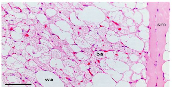
The resection specimen was partly covered by skeletal muscle and measured 40 mm, with tan-yellow and soft cut surfaces. Microscopically, the tumor consisted of brown fat cells with multivacuolated cytoplasm and univacuolated white adipocytes in equal proportions, consistent with a diagnosis of hibernoma. In the periphery, the tumor cells partly surrounded striated muscle fibers.

References
- Furlong MA, Fanburg-Smith JC, Miettinen M. The morphologic spectrum of hibernoma: A clinicopathologic study of 170 cases. Am. J. Surg. Pathol. 2001; 25: 809-814.
- Greenbaum A, Coffman B, Rajput A. Hibernoma: Diagnostic and surgical considerations of a rare benign tumour. BMJ Case Rep. 2016; 10-13.
- Sachpekidis C, Roumia S, Schwarzbach M, Dimitrakopoulou-Strauss A. Dynamic 18 F-fluorodeoxyglucose positron emission tomography/CT in hibernoma: Enhanced tracer uptake mimicking liposarcoma. World J. Radiol. 2013; 5: 498.
- Park JH, et al. The Values and Limitations of FDG-PET/CT for Diagnosis of Hibernoma. Case Rep. Orthop. 2015. doi:10.1155/2015/958690.
- Kim JD, Lee HW. Hibernoma: Intense Uptake on F18-FDG PET/CT. Nucl. Med. Mol. Imaging. 2010. doi:10.1007/s13139-012-0150-z.
- Purohit BS, et al. FDG-PET/CT pitfalls in oncological head and neck imaging. Insights Imaging. 2014; 5: 585-602.
- Smith CS, Teruya-Feldstein J, Caravelli JF, Yeung HW. False-positive findings on18F-FDG PET/CT: Differentiation of hibernoma and malignant fatty tumor on the basis of fluctuating standardized uptake values. Am. J. Roentgenol. 2008; 190: 1091-1096.
- Chen KY, et al. Brown Adipose Reporting Criteria in Imaging STudies (BARCIST 1.0): Recommendations for Standardized FDG-PET/CT Experiments in Humans. Cell Metab. 2016; 24: 210-222.