
Journal of Clinical Images and Medical Case Reports
ISSN 2766-7820
Clinical Image - Open Access, Volume 6
An incidental imaging of basilar artery fenestration
Enes Yılmaz1*; Büşra Şahin Toprak1; Furkan Akman1; Mehmet Faik Özveren2
1Department of Radiology, Medical Faculty, Kutahya Health Sciences University, Kutahya, Turkey.
2Department of Neurosurgery, Kutahya Anadolu Hospital, Kutahya, Turkey.
*Corresponding Author : Enes Yılmaz
Department of Radiology, Medical Faculty, Kutahya Health Sciences University, Kutahya, Turkey.
Tel: +905383854161;
Email: enesyilmazdr@gmail.com
Received : Apr 12, 2025
Accepted : Apr 30, 2025
Published : May 07, 2025
Archived : www.jcimcr.org
Copyright : © Yılmaz E (2025).
Keywords: Magnetic resonance imaging; Fenestration; Ischemia; Angiography.
Citation: Yılmaz E, Şahin Toprak B, Akman F, Faik Özveren M. An incidental imaging of basilar artery fenestration. J Clin Images Med Case Rep. 2025; 6(5): 3581.
Description
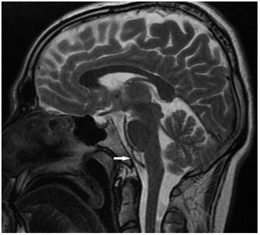
A 30-year-old male patient presented to the neurosurgery outpatient clinic with complaints of headache and neck stiffness. Non-contrast brain Magnetic Resonance Imaging (MRI) revealed a basilar artery fenestration in the prepontine region (Figure 1). Fenestration of the cerebral arteries is relatively rare; however, it is most frequently observed in the basilar artery. While the majority of cases are asymptomatic, several reports in the literature have described associations with cerebral ischemia. Among the proposed mechanisms underlying ischemia, the most widely accepted theory posits that flow disturbances caused by the fenestration may predispose individuals to cerebral ischemic events. The prevalence of basilar artery fenestration has been reported as 2.4% in computed tomography angiography and 1.2% in magnetic resonance angiography, establishing these modalities as the primary tools for detection [1]. Although fenestrations are not uncommon, to the best of our knowledge, no cases have been documented in the literature based solely on findings from conventional MRI [2,3]. In conclusion, even when discovered incidentally, close monitoring of such cases may be valuable for assessing potential long-term outcomes.
Declarations
Declaration of conflicting interests: The author(s) declared no potential conflicts of interest with respect to the research, author-ship, and/or publication of this article.
Funding: The author(s) received no financial support for the research, author-ship, and/or publication of this article.
Informed consent: We declare that written informed consent for the publication of patient information and images was provided by the patient’s legal representative.
Author’s contribution: Mehmet Faik Özveren (Writing-review & editing [lead]) and Enes Yılmaz (Writing-review & editing [supporting]) and Büşra Şahin Toprak (Writing-review & editing [supporting]) and Furkan Akman (Writing-review & editing [supporting]).
References
- Wu X, Lin A, Zhu J, Cai B. Basilar artery fenestration: an unusual possible cause of ischemic stroke? BMJ Case Rep. 2018; 2018: 2017222910. doi:10.1136/bcr-2017-222910.
- Bharatha A, Aviv R, White J, Fox A, Symons S. Intracranial arterial fenestrations: frequency on CT angiography and association with other vascular lesions. Surg Radial Anat. 2008; 30(5): 397-401. doi:10.1007/s00276-008-0340-7.
- Arraes-Aybar L, Villar-Martín A, Poyatos-Ruipérez C, Rodríguez-Boto G, Arrazola-García J. Prevalence of basilar artery fenestration by magnetic resonance angiography: A transversal study. Surg Radial Anat. 2012; 35(6): 487-93. doi:10.1007/s00276-012-1053-5.