
Journal of Clinical Images and Medical Case Reports
ISSN 2766-7820
Case Report - Open Access, Volume 6
Out of place: A Jejunal Meckel’s diverticulum in a nonverbal adolescent
*Corresponding Author : Berkay Tek Kanat
Department of Pediatric Surgery, Eskisehir City Hospital, Eskişehir 26040, Turkey.
Tel: +905374244012,
Email: dr.berkaytekkanat@gmail.com
Received : Apr 17, 2025
Accepted : May 06, 2025
Published : May 13, 2025
Archived : www.jcimcr.org
Copyright : © Tekkanat B (2025).
Abstract
Meckel’s diverticulum is the most common congenital anomaly of the gastrointestinal tract, usually located in the ileum, and often remains asymptomatic. However, it may rarely be encountered in atypical locations and present with symptoms mimicking acute abdomen. We present a case of a 14-year-old male with mental retardation who was admitted with non-specific restlessness. Due to agitation, imaging was limited and the initial diagnosis was acute appendicitis. Intraoperative exploration revealed a perforated omphalomesenteric duct remnant located in the jejunum. Segmental jejunal resection and primary anastomosis were performed. The final pathology confirmed the presence of Meckel’s diverticulum containing ectopic gastric and pancreatic tissue. This case emphasizes the need to consider atypical localizations of Meckel’s diverticulum in the differential diagnosis of acute abdomen, particularly in nonverbal patients with limited diagnostic cooperation.
Keywords: Meckel’s diverticulum; Jejunum; Omphalomesenteric duct; Acute abdomen; Nonverbal patient.
Citation: TekKanat B. Out of place: A Jejunal Meckel’s diverticulum in a nonverbal adolescent. J Clin Images Med Case Rep. 2025; 6(5): 3588.
Introduction
The omphalomesenteric duct remnant is a congenital anomaly resulting from the failure of involution of the embryonic connection between the yolk sac and the midgut. It may present in various forms, including Meckel’s diverticulum, omphalomesenteric cyst, umbilical fistula, fibrous band, or umbilical polyp [1]. Although it is the most common congenital anomaly of the gastrointestinal tract, it often remains clinically silent [2]. Meckel’s diverticulum, the most frequent form of omphalomesenteric duct remnants, contains all layers of the intestinal wall and may include ectopic mucosa [3]. It is typically located on the antimesenteric border of the ileum, approximately 7-200 cm proximal to the ileocecal valve [4]. While often asymptomatic, it can present with various complications including bleeding, obstruction, or inflammation [5]. Herein, we present a rare case of an omphalomesenteric duct remnant located in the jejunum, outlining the diagnostic challenges and surgical management in a nonverbal adolescent patient.
Case presentation
A 14-year-old male patient with a history of mental retardation was brought to the emergency department with restlessness persisting for four days. Due to the patient’s inability to communicate and express his symptoms, the family could not identify the cause of distress, resulting in delayed medical attention. The patient’s agitation, fear of unfamiliar individuals, and claustrophobia limited the possibility of conducting a thorough physical examination or abdominal Computed Tomography (CT). A bedside abdominal ultrasound was therefore performed with suspicion of acute abdomen.
Diagnosis and treatment
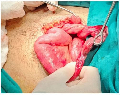
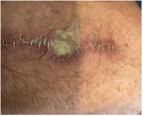
Ultrasound imaging revealed a 2,5 cm non-compressible, blind-ending tubular structure in the right lower quadrant, which was interpreted as an enlarged appendix. Based on this preliminary diagnosis, the patient was taken to the operating room for an appendectomy. A transverse incision was made in the right lower quadrant, and intraoperative exploration revealed a necrotic and perforated tubular structure with intestinal contents leaking through the perforation. Due to poor mobilization and limited access, the exploration was converted to a midline laparotomy. A further exploration revealed a necrotic and perforated omphalomesenteric duct remnant located approximately 20 cm distal to the ligament of Treitz, extending between the umbilicus and the small intestine (Figure 1). To avoid recurrence and ensure complete removal, segmental resection of the jejunum was performed, followed by end-to-end anastomosis. Enteral nutrition was initiated on postoperative day 5. Although the patient had no systemic signs of infection, a purulent discharge was noted from the surgical site (Figure 2). As laboratory findings showed no leukocytosis or elevated inflammatory markers, an infectious etiology was not suspected. Due to persistent drainage, the skin sutures were removed under sterile conditions in the operating room. The source of drainage was confirmed to be superficial, and the abdominal cavity was not entered. Local debridement was performed and the procedure was concluded. The patient was discharged in good condition on postoperative day 20. Histopathological examination revealed ectopic gastric and pancreatic mucosa consistent with Meckel’s diverticulum.
Discussion/conclusion
Among omphalomesenteric duct anomalies, Meckel’s diverticulum is the most commonly encountered, with an incidence ranging from 0.3% to 2.9% in autopsy series [4]. Despite being the most frequent congenital anomaly of the gastrointestinal tract, it is commonly asymptomatic [2]. The classical “rule of twos” is often used to describe Meckel’s diverticulum: it occurs in 2% of the population, is twice as common in males, is approximately 2 inches in length, located 2 feet from the ileocecal valve, and contains two types of ectopic tissue—most commonly gastric and pancreatic mucosa [6]. While it is typically found in the ileum, Meckel’s diverticulum may also be located in unusual sites, such as the jejunum [7]. Approximately 75% of symptomatic patients are younger than 10 years of age, with a mean age of 3.5 years [1]. Presenting symptoms may include gastrointestinal bleeding, intussusception, bowel obstruction, or perforation [7]. Diagnostic tools include abdominal ultrasound, CT scan, and technetium-99m pertechnetate scintigraphy [3]. The primary goal of surgical treatment is to completely excise the diverticulum and prevent complications related to residual ectopic mucosa. Segmental resection with primary anastomosis is preferred when ectopic tissue is suspected or confirmed [7]. In this case, the patient’s inability to communicate due to cognitive impairment led to diagnostic delays. Ultrasound findings were misinterpreted as acute appendicitis. Intraoperative findings confirmed a jejunal omphalomesenteric duct remnant with perforation, an atypical but important differential consideration. Meckel’s diverticulum, while frequently asymptomatic, can present with various clinical manifestations depending on its location and associated complications. Although classically situated in the ileum proximal to the ileocecal valve, rare localizations such as the jejunum must be considered, particularly in cases of unexplained acute abdomen. Surgical intervention remains the cornerstone of treatment to eliminate symptoms and reduce the risk of future complications through complete resection of the affected segment.
References
- Boonthai A, Mullassery D, Losty PD. Omphalomesenteric Duct Remnants. In: Puri P, Höllwarth ME, editors. Pediatric Surgery: Diagnosis and Management. Cham: Springer International Publishing. 2023; 543-50.
- Durakbasa CU, Okur H, Mutus HM, Bas A, Ozen MA, Sehiralti V, et al. Symptomatic omphalomesenteric duct remnants in children. Pediatr Int. 2010; 52(3): 480-4.
- Shaikh FA, KA DV, Shaikh H, Oduoye MO. Recognizing perforated Meckel’s diverticulum: A crucial differential in acute appendicitis imitation. Clinical Case Reports. 2024; 12(9): 9361.
- Hansen CC, Soreide K. Systematic review of epidemiology, presentation, and management of Meckel’s diverticulum in the 21st century. Medicine (Baltimore). 2018; 97(35): 12154.
- Ponniah K, Kinsey-Trotman S, Agarwal N, Lachlan D, Kozman M. Enteric Duplication Cyst Associated With Meckel’s Diverticulum: A Rare Cause of Acute Abdomen. Journal of Investigative Medicine High Impact Case Reports. 2024; 12: 23247096241271986.
- Ahmed M, Saeed R, Allawi A, Zajicek J. Meckel’s Diverticulum With Perforation. Cureus. 2024.
- Permekerlis A, Varvara ST, Gemousakaki E, Tepelidis C, Fotiadis P. Acute Abdomen Accompanied by Torsion of a Segment of the Jejunum Due to Meckel’s Diverticulum: A Rare and Interesting Case. Cureus. 2024.