Journal of Clinical Images and Medical Case Reports
ISSN 2766-7820
Clinical Image - Open Access, Volume 6
The CT and MRI in the non coronary sinus aneurysm
Caran Facundo Yamil1*; Garrido Mariel Desiree1; Pérez Ferro José1; Bayol Pablo Augusto1; Collantes José2; Aguirre Pablo3
1Department of Radiology, Instituto de Cardiología de Corrientes “Juana Francisca Cabral”, Cordoba 1259, Corrientes Capital, Argentina.
2Department of of Surgery, Instituto de Cardiología de Corrientes “Juana Francisca Cabral”, Cordoba 1259, Corrientes Capital, Argentina.
3Inpatient Department, Instituto de Cardiología de Corrientes “Juana Francisca Cabral”, Cordoba 1259, Corrientes Capital, Argentina.
*Corresponding Author : Caran Facundo Yamil
Department of Radiology, Instituto de Cardiología
de Corrientes “Juana Francisca Cabral”, Cordoba
1259, Corrientes Capital, Argentina.
Tel: +54-3772465841;
Email: facucaran@gmail.com
Received : Apr 30, 2025
Accepted : May 20, 2025
Published : May 27, 2025
Archived : www.jcimcr.org
Copyright : © Yamil CF (2025).
Citation: Yamil CF, Desiree GM, José PF, Augusto BP, José C, et al. The CT and MRI in the non coronary sinus aneurysm. J Clin Images Med Case Rep. 2025; 6(5): 3608.
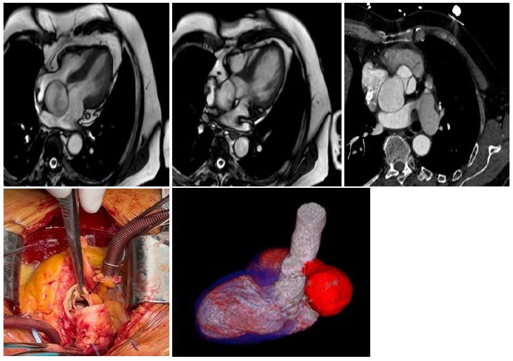
Description
Coronary sinus aneurysms are a rare entity, usually congenital, although they can be associated with conditions such as infective endocarditis and Behçet’s disease. They most frequently originate in the right coronary sinus (70%–90%) and rarely in the non-coronary sinus and may be asymptomatic until complications develop. Complications include rupture, arrhythmias, outflow tract obstruction, and aortogenic embolism. Surgical treatment is the method of choice, with repair by direct closure or prosthetic patch placement depending on the size of the aneurysm. We present a case of a non-coronary sinus aneurysm in a 78-year-old male patient, diagnosed by cardiac magnetic resonance imaging (MRI) and cardiac computed tomography (CT) angiography. The patient was treated with Dacron patch aortoplasty, with good clinical outcome. This case highlights the importance of imaging methods in the diagnosis and treatment of coronary sinus aneurysms, a rare but potentially serious condition.