
Journal of Clinical Images and Medical Case Reports
ISSN 2766-7820
Case Report - Open Access, Volume 2
Myelodysplastic syndrome: Discordant involvement of the marrow as observed in the bone marrow biopsy sections
Anwarul Islam
Clinical Associate Professor of Medicine, Division of Hematology/Oncology, Department of Medicine, Buffalo General Hospital, Room E 318, Buffalo, New York 14203, USA
*Corresponding Author : Anwarul Islam
Clinical Associate Professor of Medicine, Division of
Hematology/Oncology, Department of Medicine, Buffalo General Hospital, Room E 318, Buffalo, New York
14203, USA
Email: aislam@buffalo.edu
Received : Feb 08, 2021
Accepted : Mar 15, 2021
Published : Mar 17, 2021
Archived : www.jcimcr.org
Copyright : © Islam A (2021).
Clinical image description
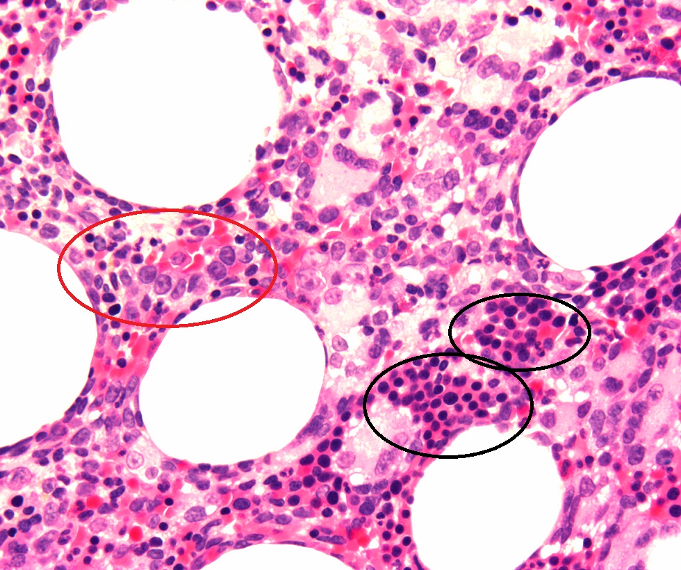
Myelodysplastic syndromes (MDS) are a diverse group of hematological disorders that affect the blood and bone marrow. Laboratory diagnosis of MDS is almost exclusively based on the morphology of hematopoietic cells observed in smears of peripheral blood and bone marrow and this depends solely on the presence of dysplastic changes observed in the granuloid, erythroid, and megakaryocytic cells [1,2]. Up until now evaluation of sections of bone marrow trephine biopsy specimen did not play a significant role in its diagnosis. Bone marrow is not always involved uniformly in pathological processes and morphological discordance is well known [3,4]. This is particularly true for patients with MDS. We present a case of 75 years old white male with a diagnosis of MDS. His bone marrow biopsy section showed areas of clusters of immature myeloid precursor cells (Figure 1 - red circle) and areas of clusters of immature erythroid precursor cells (Figure 1 - black circles) in different areas of the same section admixed with mature hematopoietic cells.
It is postulated that during bone marrow aspiration if the tip of the aspirating needle encountered one of the areas of clusters of immature myeloid precursor cells (Figure 1-red circle) the aspirate may have contained 15-20% or more blast cells and the patient might have been misleadingly diagnosed as high risk MDS or even de novo acute myeloid leukemia. On the other hand during bone marrow aspiration if the tip of the aspirating needle missed these areas of focal collection of myeloid blast cells and struck an area of the marrow containing clusters of erythroid precursor cells (Figure 1- black circles) such a bone marrow aspirate would have few myeloblasts and more erythroid precursors and the patient might have been considered as of low-risk MDS. Alternatively, in this situation, the case could have been misleadingly diagnosed as erythroleukemia (FAB M6). It is proposed that evaluation of sections of bone marrow biopsy should be included in the diagnosis and management of patients with MDS.
Citation: Islam A. Myelodysplastic syndrome: Discordant involvement of the marrow as observed in the bone marrow biopsy sections. J Clin Images Med Case Rep. 2021; 2(2): 1029.
References
- Bennett JM, Catovsky D, Daniel MT, et al. The French- AmericanBritish (FAB) Co-operative group. Proposals for the classification of the myelodysplastic syndromes. Br J Haematol. 1982; 51: 189- 199.
- Vardiman JW, Brunning RD, Arber DA, Le Beau MM. Introduction and overview of the classification of the myeloid neoplasms. In: Swerdlow SH, Campo E, Harris NL, et al, eds. WHO Classification of Tumours of Haematopoietic and Lymphoid Tissues. 4th ed. Lyon, France: IARC Press; 2008: 18-30. WHO Classification of Tumours; vol 2.
- Islam A, Catovsky D, Goldman JM, Galton DAG. Value of Long Core Biopsy in Detection of Discrete Bone Marrow Lesions. 1979: Lancet 878.
- Islam A. A new bone marrow aspiration needle to overcome the sampling errors inherent in the technique of bone marrow aspiration. Journal of Clinical Pathology. 1983: 36; 954-958.