Journal of Clinical Images and Medical Case Reports
ISSN 2766-7820
Case Report - Open Access, Volume 2
Sebaceous gland extrusion
Francisco Javier Torres-Gómez*; Rosa Sánchez de Medina-González
Pathology Laboratory, CITADIAG SL, c/ Amor de Dios 17 a, loc B, 41002, Sevilla, Spain.
*Corresponding Author : Francisco Javier Torres Gómez
Pathology Laboratory, CITADIAG SL, c/ Amor de Dios 17 a,
loc B. 41002, Sevilla, Spain.
Email: javiertorresgomez@yahoo.es
Received : Mar 15, 2021
Accepted : Apr 23, 2021
Published : Apr 27, 2021
Archived : www.jcimcr.org
Copyright : © Torres-Gómez FJ (2021).
Citation: Torres-Gómez FJ, Medina-González RSD. Sebaceous gland extrusión. J Clin Images Med Case Rep. 2021; 2(2): 1075.
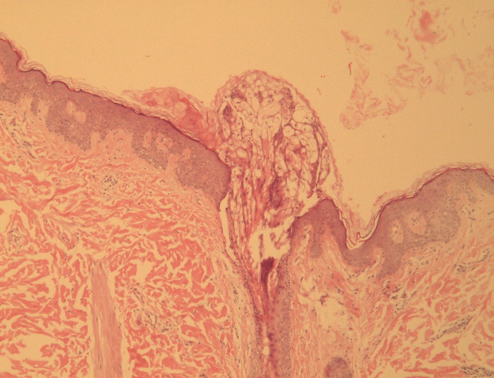
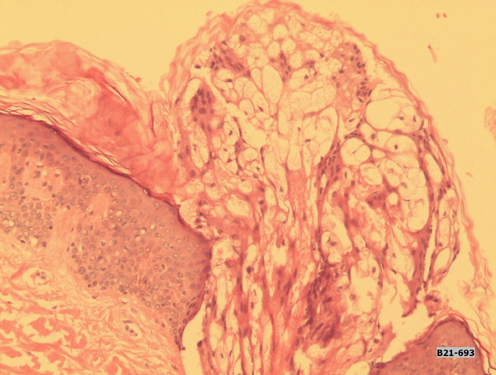
Description
The extrusion of a sebaceous gland through a follicular infundibulum is a rare dermatopathology image that raises two fundamental questions: Is it an artifact?. Can a full gland be expelled just as its secretion does?.
The answer is not easy and there are no bibliographic references to help us solving the doubt.
These two images correspond to the case of an 18-year-old male patient who consulted for a rapidly evolving yellowish nummular lesion on the cheek. An oily secretion was released from it.
The patient denied manipulation of the lesion. With the clinical judgment of sebaceous gland hyperplasia, the lesion was excised. The presence of an extruded sebaceous gland was the only histological finding (Figures 1,2). Excision was curative.