Journal of Clinical Images and Medical Case Reports
ISSN 2766-7820
Case Report - Open Access, Volume 2
Image in medicine: Cement pulmonary embolism
Asarisi F; Heme N; Fourrier E; Ferrari E*
Cardiology Department, Pasteur University Hospital, Nice, France.
*Corresponding Author : Ferrari E
Cardiology Department, Pasteur University Hospital,
30 av de la voie romaine, 06001 Nice, France.
Email: ferrari.e@chu-nice.fr
Received : Apr 07, 2021
Accepted : May 04, 2021
Published : May 07, 2021
Archived : www.jcimcr.org
Copyright : © Ferrari E (2021).
Citation: Asarisi F, Heme N, Fourrier E, Ferrari E. Image in medicine: Cement pulmonary embolism. J Clin Images Med Case Rep. 2021; 2(3): 1119.
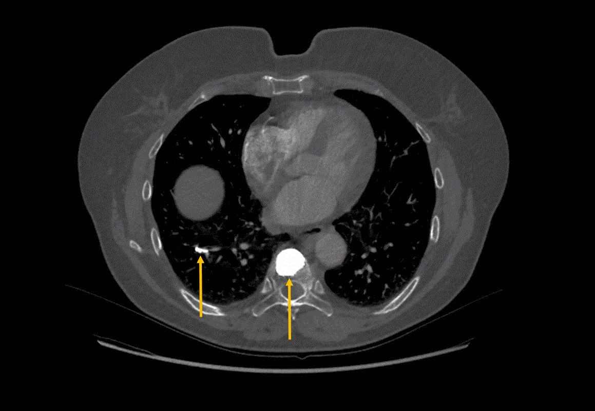
Clinical image description
A 65-year-old woman was treated for vertebroplasty (cement injection). 24 hours after the procedure, she complains chest pain. A CT scan was performed which revealed on the same image section the presence of highly radiopaque material in a right basal segmental artery the density of which is strictly identical to that of the cement of the treated vertebra (yellow arrows).
This cement pulmonary embolism canot be disolved by an anticoagulant treatment.