
Journal of Clinical Images and Medical Case Reports
ISSN 2766-7820
Case Report - Open Access, Volume 2
Right ovarian torsion and oophorectomy in second trimester of pregnancy with subsequent live birth at term
Omotade A Ijarotimi1,2*; Olumide A Adeniyi2; Stephen O Omitinde2; Akaninyene E Ubom2; Momoh Ibrahim2; Adebanjo B Adeyemi1,2
1 Department of Obstetrics, Gynaecology, and Perinatology, Faculty of Clinical Sciences, College of Health Sciences, Obafemi Awolowo University, Ile-Ife, Osun state, Nigeria.
2 Department of Obstetrics, Gynaecology, and Perinatology, Obafemi Awolowo University Teaching Hospitals Complex, Ile-Ife, Osun state, Nigeria.
*Corresponding Author: Omotade A Ijarotimi
Department of Obstetrics, Gynaecology, and Perinatology, Faculty of Clinical Sciences, College of Health Sciences,
Obafemi Awolowo University, Ile-Ife, Osun State, Nigeria.
Email: oijarotimi@cartafrica.org
Received : Jun 15, 2021
Accepted : Jul 16, 2021
Published : Jul 21, 2021
Archived : www.jcimcr.org
Copyright : © Ijarotimi Omotade A (2021).
Abstract
Introduction: Ovarian torsion is a cause of acute abdomen in pregnancy with an incidence of less than 1% occurring most commonly in the first trimester. The symptoms are non-specific with a propensity for missed or delayed diagnosis which may worsen the morbidity associated with this condition. Management is mainly surgical and pregnancy outcome is usually satisfactory.
Case presentation: We present the case of a 29-year-old G2 P0 +1 with background polycystic ovaries who complained of abdominal pains at an Estimated Gestational Age (EGA) of 21 weeks in pregnancy. Clinical suspicion was confirmed with a 2D abdominopelvic ultrasound and she subsequently had emergency exploratory laparotomy with right oophorectomy. Pregnancy progressed well and she had a live birth at term.
Conclusion: Ovarian torsion is rare in pregnancy. Complications following prompt surgical intervention are however, few and pregnancy outcomes are usually favourable.
Keywords: Ovarian cyst; torsion; acute abdomen; cyst accident; pregnancy.
Citation: Ijarotimi OA, Adeniyi OA, Omitinde SO, Ubom AE, Momoh I, Adeyemi AB. Right ovarian torsion and oophorectomy in second trimester of pregnancy with subsequent live birth at term. J Clin Images Med Case Rep. 2021; 2(4): 1239.
Introduction
The presence of ovarian cyst or mass in pregnancy is a common finding with an incidence ranging between 1 to 5% depending on the criteria used and the timing of diagnosis [1,2]. Diagnosis of ovarian cyst has been on the rise of late, with increased uptake of assisted reproductive technology and wide spread use of ultrasound in the first trimester. Most of these cysts are small (<5 cm), benign and regress as the pregnancy advances [3]. However ovarian cyst accidents including infection, hemorrhage and torsion can occur leading to an acute abdomen. The risk of ovarian torsion rises by 5 fold during pregnancy with an incidence of 5 per 10,000 pregnancies [4]. The symptoms of ovarian torsion in pregnancy are nonspecific leading to delayed diagnosis. A high index of suspicion, early and appropriate deployment of doppler ultrasound will detect ovarian torsion and may help with ovarian tissue conservation. We present the case of a women with delayed diagnosis of ovarian torsion in pregnancy who had laparotomy with right oophorectomy and subsequent live delivery at term.
Case presentation
A 29 year old G2P0+1 presented to the obstetric emergency unit at EGA of 21 weeks with complaint of lower abdominal pain of six hours duration which was colicky in nature (waxing and waning) and relieved by lying down. There was no nausea, vomiting or fever and she did not have any urinary symptoms. She had no history of trauma to the abdomen and no previous diagnosis of co-existing uterine fibroid. There was no change in bowel habit, no yellowness of the eyes or pruritus. There was no bleeding per vaginam or drainage of liquor. She had similar complaints three months earlier at EGA six weeks with ultrasound scan revealing a right adnexal cyst likely corpus luteum cyst measuring 1.96 X 1.90 cm. she was managed conservatively and discharged. She had no chronic medical condition or allergies but was diagnosed with polycystic ovary syndrome about five years prior to the index pregnancy. She did not use any ovulation inducing drug as the index pregnancy was spontaneously conceived. She does not take alcohol nor smoked cigarette.
At presentation, she was in obvious painful distress, she was not pale, febrile nor dehydrated and she had no pedal edema. Her vital signs were essentially normal with a pulse rate of 88 beats per minute, respiratory rate of 20 cycles per minute, blood pressure of 120/70 mmHg, and temperature of 36.5o C. The abdomen was uniformly enlarged (gravid) with moderate to marked tenderness in the right iliac fossa associated with guarding but not rebound tenderness. The height of fundus was approximately 20 weeks’ size and the fetal heart tone was heard, regular and asynchronous with the maternal pulse. There was difficulty in palpating or appreciating any adnexal mass because of tenderness.
A diagnosis of Acute abdomen possibly due to acute appendicitis was made to rule out ureteric colic. Results of full blood count, renal and liver function tests were normal. A pelvic scan showed a single viable fetus, the appendix wasn’t visualized and there was no comment on the adnexa. She was placed on analgesics and liberal fluids. A surgical review by the General Surgeons and Urologists supported the earlier diagnosis by the obstetric team and she was managed conservatively.
She continued to have breakthrough episodes of pain despite the analgesia and with complaints of vomiting. A repeat ultrasound scan was done about 24 hours after admission which revealed a viable fetus at 20 weeks gestational age and a markedly enlarged right ovary with heterogenous hypoechoic prominent medulla and few peripherally arranged follicles seen within it measuring 8.3 X 4.0 X 6.1 cm equivalent to 105 mls. Color flow doppler showed no significant parenchymal perfusion. There was mild free anaechoic fluid in the pelvis. The left ovary was normal (4.4 X 2.1 X 2.5 cm and volume of 12.4 mls). A diagnosis of torsion of the right ovary was made, she was counselled and informed consent obtained for emergency exploratory laparotomy.
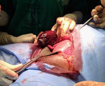
At surgery, there was a 20 weeks size gravid uterus. In addition, 100 mls of hemorrhagic fluid was found in the pelvis and a right hemorrhagic ovarian mass measuring 8 cm X 6 cm twisted clockwise three and half times (Figure 1) on its longitudinal axis was noted. There was involvement of the right fallopian tube in the torsion, the tube was initially dusky but later became well perfused after untwisting the mass. The left ovary was not visualized. She subsequently had right oophorectomy done with special care taken to avoid manipulation of the gravid uterus. She had a satisfactory postoperative recovery and was placed on analgesics, antibiotics and tocolytics. She was discharged home on the fifth post-operative day. Histology of the mass showed a greyish white tissue measuring 8.5 X 6.5 X 4.0 cm and the surface had multiple ecchymoses as well as petechiae haemorrhages. Cut surfaces showed extensively hemorrhagic tissue with multiple cysts filled with gelatinous materials. There was extensive hemorrhagic necrosis and the few surviving ovarian tissues show some dilated cysts lined by many layers of granulosa cells. No atypical cell was seen. This was consistent with ovarian torsion.
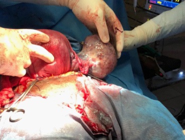
Pregnancy continued uneventfully till the gestational age of 37 weeks when she developed preeclampsia and had caesarean delivery on account of maternal request with the delivery of a live male baby weighing 3.25 kg and good Apgar scores. Intraoperatively, the left ovary was found to be multi-cystic and enlarged measuring about 8 X 6 X 2 cm (Figure 2).
Discussion
Ovarian torsion is a relatively rare event in pregnancy in which there is total or partial rotation of the adnexa around its vascular axis or pedicle and should be considered in the differential diagnosis of any woman with acute abdomen [5,6]. The incidence is reported as 0.03% and in women presenting to the early pregnancy unit in the 1st trimester with identified ovarian cyst during routine scan; the rate of torsion is about 3%. The risk of torsion increases by 15-fold once a cyst has been identified in early pregnancy [7]. While most ovarian torsion are associated with the presence of an ovarian cyst, torsion can occur in previously normal adnexa [8,9].
The exact reason why the incidence of ovarian torsion is increased during pregnancy is unknown but may be as a result of the movement of the adnexa with an associated cyst out of the pelvis giving space for rotation. Torsion has been reported in all trimesters of pregnancy, but it is more common within the first trimester. The probable explanation for this is the higher incidence of functional cysts in the first trimester most of which will regress as the pregnancy proceeds with only about <5% persisting [10,11]. The index case had persistent ovarian cyst with torsion in the second trimester of pregnancy. Other risk factors for ovarian torsion include use of assisted reproduction techniques especially with ovulation induction or ovarian hyperstimulation. Torsion in the Presence of Polycystic Ovaries (PCOS) is also reported to be increased either as a result of need for ovulation induction or the size and volume of the ovary itself [5,6].
The right ovary is more commonly involved in torsion than the left. It has been postulated that the sigmoid colon helps prevent torsion of the left-sided adnexa and also the likelihood of surgeons to perform surgical exploration for right-sided abdominal pain to rule out acute appendicitis [5,9,12]. Common presenting features are not different from non-pregnant women and include sudden onset abdominal pain, nausea and or vomiting and adnexal mass [8,12]. However, these symptoms are nonspecific and diagnoses such as appendicitis, pyelonephritis, degenerating uterine fibroids, ureteric colics or even preterm labour are often entertained. Between 15-30% of ovarian torsion are misdiagnosed or delayed in the second and third trimester of pregnancy because the increased size of the uterus causes some difficulties in abdominal palpation and may impede efficient sonographic examination [5]. This was the case in the first ultrasound scan done at presentation by this patient. Missed or delayed diagnosis has implications on the management modality, ability to preserve ovarian tissue and the reproductive potential of the woman.
Ultrasound is usually the first investigation of choice for diagnosis with a sensitivity of 71% and doppler interrogation will reveal reduced or absent blood flow to the ovary [5,9]. Other imaging modalities such as CT scan and MRI are rarely indicated but maybe useful when the diagnosis is in doubt and the risk of malignancy is elevated. Management is mainly surgical which could be via laparotomy or laparoscopy. Despite the safety of laparoscopy and the reports that the procedure can be carried out in the third trimester, pregnant women are more likely to undergo laparotomy when compared with their non-pregnant counterpart [3,4]. De-torsion with removal of the offending cyst and subsequent oophoropexy has been reported where the diagnosis and intervention is made early. However, torsion is traditionally managed with oophorectomy where the ovary appeared engorged or ischaemic as in this case. Salpingo-oophorectomy is done if the fallopian tube is involved in the ischaemic necrosis [13].
Postoperative complications are few and pregnancy outcomes following surgical intervention for ovarian torsion are usually favourable with most of the pregnancies carried to term [5].
Conclusion
Ovarian torsion is rare in pregnancy and the diagnosis is often missed due to nonspecific clinical features. With a high index of suspicion, affected women can be treated immediately to preserve ovarian function. Complications following prompt surgical intervention are few and pregnancy outcomes are usually favourable.
Acknowledgements: IOA was supported by the Consortium for Advanced Research Training in Africa (CARTA). CARTA is jointly led by the African Population and Health Research Center and the University of the Witwatersrand and funded by the Carnegie Corporation of New York (Grant No–B 8606.RO2), Sida (Grant No:54100029), the DELTAS Africa Initiative (Grant No: 107768/Z/15/Z). The DELTAS Africa initiative is an independent funding scheme of the African Academy of Sciences (AAS)’s Alliance for Accelerating Excellence in Science in Africa (AESA) and supported by the New Partnership for Africa's Development Planning and Coordinating Agency (NEPAD Agency) with funding from the Wellcome Trust (UK) and the UK government. The statements made and views expressed are solely the responsibility of the Fellow.
Authors' information
IOA: MBChB, MPH, FMCOG (Nig.), CARTA PhD fellow, Senior Lecturer, Consultant Obstetrician and Gynaecologist.
AOA: MBBS, FWACS, Consultant Obstetrician and Gynaecologist.
OSO: MBBS, FWACS, Consultant Obstetrician and Gynaecologist.
UAE: MBBS, MMCOG, MWACS, Senior Specialist Registrar.
MI: MBBS, MMCOG, MWACS, Senior Specialist Registrar.
AAB: MBBS, FWACS, Professor of Obstetrics and Gynaecology.
References
- Eidenberger-Gautshi T, Smith A, Sayasneh A. Ovarian masses in pregnancy: a single centre retrospective study. Br J Med Pract. 2018; 11: 1109.
- Mukhopadhyay A, Shinde A, Naik R. Ovarian cysts and cancer in pregnancy. Best Pract Res Clin Obstet Gynaecol. 2016; 33: 58-72.
- Chen L, Ding J, Hua K. Comparative analysis of laparoscopy versus laparotomy in the management of ovarian cyst during pregnancy. J Obstet Gynaecol Res. 2014; 40: 763-769.
- Bassi A, Czuzoj-Shulman N, Abenhaim HA. Effect of Pregnancy on the Management and Outcomes of Ovarian Torsion: A Population-Based Matched Cohort Study. J Minim Invasive Gynecol. 2018; 25: 1260-1265.
- Chang SD, Yen CF, Lo LM, Lee CL, Liang CC. Surgical intervention for maternal ovarian torsion in pregnancy. Taiwan J Obstet Gynecol. 2011; 50: 458-462.
- Erdemoglu M, Kuyumcuoglu U, Kale A. Pregnancy and adnexal torsion: analysis of 20 cases. Clin Exp Obstet Gynecol. 2010; 37: 224-225.
- Asfour V, Varma R, Menon P. Clinical risk factors for ovarian torsion. J Obstet Gynaecol. 2015; 35: 721-725.
- Melcer Y, Sarig-Meth T, Maymon R, Pansky M, Vaknin Z, et al. Similar But Different: A Comparison of Adnexal Torsion in Pediatric, Adolescent, and Pregnant and Reproductive-Age Women. J Womens Health (Larchmt). 2016; 25: 391-396.
- Bottomley C, Bourne T. Diagnosis and management of ovarian cyst accidents. Best Pract Res Clin Obstet Gynaecol. 2009; 23: 711-724.
- Brady PC, Simpson LL, Lewin SN, Smok D, Lerner JP, et al. Safety of conservative management of ovarian masses during pregnancy. J Reprod Med. 2013; 58: 377-382.
- Goh WA, Rincon M, Bohrer J, Tolosa JE, Sohaey R, Riano R, et al. Persistent ovarian masses and pregnancy outcomes. J Matern Fetal Neonatal Med. 2013; 26: 1090-1093.
- Senarath S, Ades A, Nanayakkara P. Ovarian cysts in pregnancy: a narrative review. J Obstet Gynaecol. 2020: 1-7.
- Hosny TA. Oophoropexy for ovarian torsion: A new easier technique. Gynecol Surg. 2017; 14: 7.