
Journal of Clinical Images and Medical Case Reports
ISSN 2766-7820
Case Report - Open Access, Volume 2
Cysticercotic fourth ventricle ependymitis
Carlos Cosentino1,2*; Miriam Vélez1; Luis Torres1
1 Department of Neurodegenerative Disorders, Instituto Nacional de Ciencias Neurológicas, Peru.
2 School of Medicine of Universidad Nacional Mayor de San Marcos, Peru.
*Corresponding Author: Carlos Cosentino
Department of Neurodegenerative Disorders,
Instituto Nacional de Ciencias Neurológicas, Ancash
1271, Lima 1, Peru.
Email: ccosentinoe@gmail.com
Received : May 03, 2021
Accepted : Sep 27, 2021
Published : Oct 04, 2021
Archived : www.jcimcr.org
Copyright : © Cosentino C (2021).
Abstract
Neurocysticercosis is endemic in many regions of the world. Extra parenchymal forms of neurocysticercosis are infrequent but may be associated with hydrocephalus. We report the case and show a typical MRI of fourth ventricle ependymitis caused by cysticercosis.
Keywords: ependymitis; cysticercosis.
Citation: Cosentino C, Vélez M, Torres L. Cysticercotic fourth ventricle ependymitis. J Clin Images Med Case Rep. 2021; 2(5): 1339.
Introduction
Nuerocysticercosis is the infection of the CNS and the meninges by the larval stage of the Taenia solium. Neurocysticercosis is endemic in most Latin American countries where is the major cause of adult-onset epilepsy.
Case report
A 16 year-old man had progressive headache, nausea, vomiting, horizontal diplopia and visual failure for two weeks. On examination bilateral horizontal nystagmus, papilledema, and multidirectional diplopia were found.
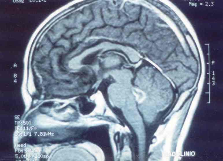
A CT scan showed obstructive hydrocephalus and contrast enhancement of fourth ventricle walls and a small right frontal calcified lesion. Western Blot for cysticercosis was positive in serum and in CSF. Ventriculoperitoneal shunt was performed with marked clinical improvement. Three weeks later an MRI showed aqueduct stenosis with partial collapse of fourth ventricle with enhanced walls because of cysticercosis ventriculitis [1] (Figure).
Discussion
The clinical expression, management and prognosis of neurocysticercosis vary depending of the number, size, stage and location of lesions as well as the presence or absence of inflammatory response of the host [2].
Patients with intraparenchymal cerebral parasites may be asymptomatic or present with seizures. Patients with intraventricular cysts and/or basal subarachnoid cysticerci cause hydrocephalus and/or intracranial hypertension and are associated with a large burden of morbidity and significant mortality. Fourth ventricle ependymitis is a very rare complication of neurocysticercosis.
Conclusions
Even if intraventricular cysts may be seen on CT as hypodense lesions that distort the ventricles they are by far better visualized on MRI as single or multiple cysts more frequent in the lateral ventricules or in the fourth ventricle.
MRI is the best accurate technique to assess the magnitude of hyperintense signals in ependymal layers.
References
- García HH, Nash TE, Del Brutto OH. Clinical symptoms, diagnosis and treatment of neurocysticercosis. Lancet Neurol 2014; 13: 1205-1215
- Del Brutto OH, Garcia HH. Neurocysticercosis. Handb Clin Neurol. 2013; 114: 313-25.