
Journal of Clinical Images and Medical Case Reports
ISSN 2766-7820
Case Report - Open Access, Volume 2
Lens coloboma secondary to neglected buphthalmos
Mousumi Banerjee
Dr Rajendra Prasad Centre for Ophthalmic Sciences, AIIMS, Delhi, India.
*Corresponding Author: Mousumi Banerjee
Dr Rajendra Prasad Centre for Ophthalmic Sciences,
AIIMS, Delhi, India.
Email: banerjeemou12@gmail.com
Received : Sep 09, 2021
Accepted : Oct 12, 2021
Published : Oct 19, 2021
Archived : www.jcimcr.org
Copyright : © Banerjee M (2021).
Citation: Banerjee M. Lens coloboma secondary to neglected buphthalmos. J Clin Images Med Case Rep. 2021; 2(5): 1370.
Clinical image description
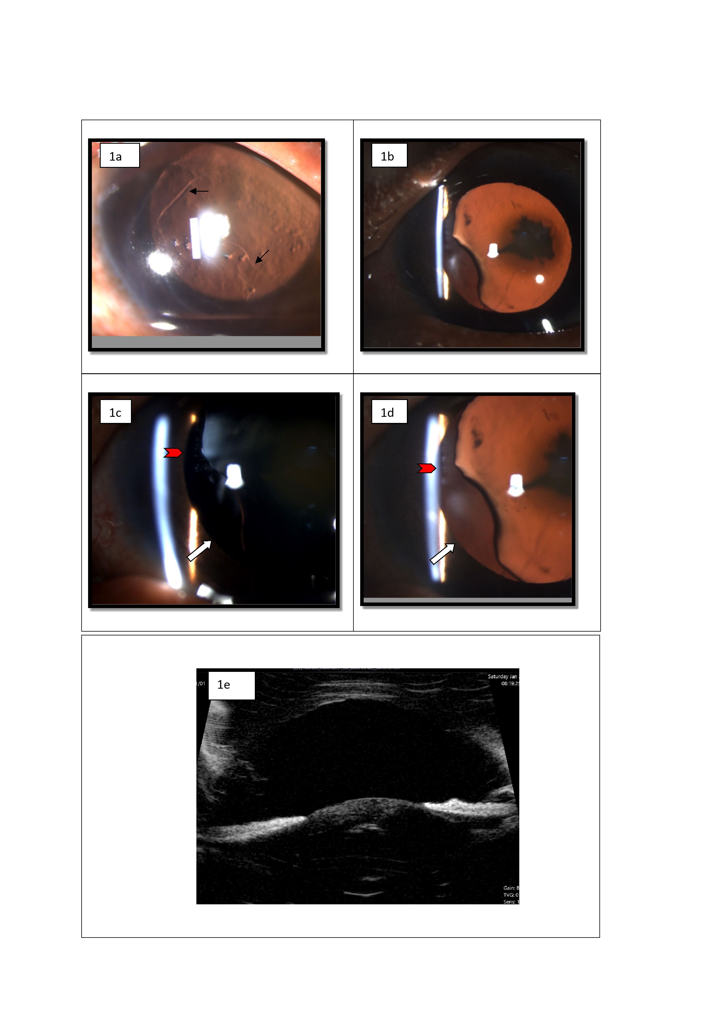
A 36-year-old male presented with progressive diminution of vision OS for 3 months. He was a known case of bilateral congenital glaucoma who underwent trabeculectomy at 6 months and 5 years of age OD and OS respectively. Best corrected visual acuity was 6/24 OD and 1/60 OS. Slit lamp examination revealed bilateral Haab striae (Figure 1a) with lens subluxation OS evident by the scalloped border of the lens with broken zonules in the superonasal quadrant and lens coloboma in the inferonasal quadrant with the absence of zonules (Figure 1b,c,d). A posterior subcapsular cataract was also noted OS. Advanced glaucomatous optic nerve cupping was noted OU. An intraocular pressure of 10 mm Hg OD and 16 mm Hg OS was noted. Biometry documented an axial length of 26.30 mm OD and 28.75 mm OS with a keratometry of 42.50D/46.50D @20˚/110˚OD and 37.75D/40.00D @ 45˚/135˚OS. Ultrasound bio-microscopy depicted increased sphericity of the lens with broken zonules OS (Figure 1e).
Lens coloboma is a misnomer as it does not indicate actual absence or defect in lenticular substance. It develops due to absence of zonules which lead to localized loss of tension on lens capsule, resulting in retraction of the lens and formation of a notch or fissure [1,2]. It is technically a coloboma of the zonule and not of the lens [3]. Lens coloboma is a congenital anomaly associated with ocular anomalies including iris, choroid or optic disc coloboma [4] or might be one of the manifestations of systemic diseases, including Marfan syndrome and Marshall syndrome [5]. In this case, coloboma is more likely to be an acquired condition secondary to uncontrolled globe enlargement OS as evident by the neglected buphthalmos till 5 years of age leading to excessive zonular stretching. Zonular discontinuity led to the scalloped border of the lens superonasally and absence of zonules inferonasally led to flattening of the lens giving a coloboma like appearance. Thus, lens coloboma can also be a sign of uncontrolled primary congenital glaucoma apart from extreme buphthlamos. Routine ophthalmic screening is required in such cases to prevent lenticular astigmatism and subsequent amblyopia.
Learning points
1. Lens coloboma can be a sign of uncontrolled primary congenital glaucoma.
2. Uncontrolled enlargement of the globe secondary to neglected glaucoma can lead to excessive zonular stretching
and formation of lens coloboma.
3. Surgical treatment is required if coloboma causes significant lenticular astigmatism leading to amblyopia, associated cataract or if the lens edge bisects the pupil.
References
- Agarwal T, Saxena R, Vajpayee R. Ultrasound biomicroscopy in lens “coloboma”. Eur J Ophthalmol. 2003; 13: 390–1.
- Duke-Elder S. Anomalies of the lens. In: Duke-Elder S, ed. Congenital Deformities, Normal and Abnormal Development (System of Ophthalmology, Vol 3). London: Henry Kimpton. 1964: 706–8.
- Onwochei BC, Simon JW, Bateman JB, Couture KC, Mir E. Ocular colobomata. Surv Ophthalmol. 2000; 45: 175-94.
- Bavbek T, Ogüt MS, Kazokoglu H. Congenital lens coloboma and associated pathologies. Doc Ophthalmol. 1993; 83: 313-22.
- Mehrotra AS, Solanki N, Sabharwal KK. Bilateral coloboma of lens in Marfan’s syndrome. Indian J Ophthalmol. 1985; 33: 201- 2.