
Journal of Clinical Images and Medical Case Reports
ISSN 2766-7820
Case Report - Open Access, Volume 2
Tuberculous peritonitis during pregnancy: Case report
Ounci Es-saad1*; Saloua Tanouti2; Houssam Bkiyar1; Ahmed Mimouni2; Brahim Housni1
1 Gynecology and Obstetrics, Faculty of Medicine Oujda, Mohammed VI University Hospital Oujda, Mohammed First University Oujda, Morocco
2 Anesthesiology and Critical Cares, Faculty of Medicine Oujda, Mohammed VI University Hospital Oujda, Mohammed First University Oujda, Morocco.
*Corresponding Author: Ounci Es-saad
Anesthesiology and Critical Cares Department,
Mohammed VI University Hospital Oujda, Morocco,
Faculty of Medicine Oujda, Morocco, Mohammed First
University Oujda, Morocco.
Email: ouncilfen@gmail.com
Received : Oct 22, 2021
Accepted : Dec 07, 2021
Published : Dec 14, 2021
Archived : www.jcimcr.org
Copyright : © Es-saad O (2021).
Abstract
Introduction: Tuberculous Peritonitis (TP) in pregnancy is a rare condition, and the diagnosis can be deleyed because of the symptoms non specificity.
Case report: 31-year-old parturient primigravidia at 34 weeks of amenorrhea presented diffuse and intense abdominal pain with vomiting. Examination found abdominal distention with ascites. Investigations did not lead to a precise diagnosis, therefore, an exploration by laporotomy with cesarean delivery were performed. The diagnosis of Tuberculous peritonitis on histopathologic examination was confirmed.
Discussion: Tuberculous peritonitisis extremely rare in pregnancy, and the rate of TP among all forms of tuberculosis varies from 0.1% to 0.7% worldwide. Gold standards for the diagnosis of TP are the identification of M. tuberculosis in ascites and peritoneal biopsy findings compatible with tuberculosis. Bacteriologic examination of the biopsy specimen should be performed, because this could be positive for tuberculosis when histological examination is negative.
Conclusion: The early diagnosis and treatment of TP in pregnancy are important to prevent obstetric and neonatal morbidities.
Keywords: tuberculous peritinitis; pregnancy; rare; delayed diagnosis.
Abbreviations: TP: Tuberculous Peritonitis; ICU: Intensive Care Unit; AST: Aspartate Aminotransferase; ALT: Alanine Transaminase.
Citation: Es-saad O, Tanouti S, Bkiyar H, Mimouni A, Housni B, et al. Tuberculous peritonitis during pregnancy: Case report. J Clin Images Med Case Rep. 2021; 2(6): 1478.
Introduction
Tuberculous peritonitis (TP) in pregnancy is a rare form of extra pulmonary tuberculosis that is not easily diagnosed of the diagnosis can be delayed because the results of radiologic evaluations and laboratory investigations are usually non-specific [1]. The early course of the disease has fewer manifestations, resulting in late presentation with grave complications like infertility and ectopic pregnancy. Surgical exploration remains essential for a diagnosis of certainty. The early diagnosis and treatment of TP in pregnancy are important to prevent obstetric and neonatal morbidities.
Case report
We report the case of a 31-year-old parturient primigravidia, the gestational age of the current pregnancy was estimated at 34 weeks of amenorrhea. This pregnancy was well monitored, with tow ultrasounds performed at 12 weeks, and 22 weeks of amenorrhea. The patient was kept in hospital for vomiting at 24 weeks of amenorrhea and was discharged after normal investigations with symptomatic treatment.
The parturient was re-admitted to the emergency room for diffuse and intense abdominal pain with vomiting.
On admission, physical examination revealed a conscious patient who was instable hemodynamically with a blood pressure of 90 mmhg/50 mmhg and a heart rate of 120 bpm, a respiratory rate of 25 cycle per minute and body temperature 38.3°C. There was signs of dehydration with unquantified weight loss.
Gyneco-obstetrical examination revealed diffuse abdominal tenderness with abdominal distention. Fetal heart sounds were perceived, and no uterine contracture was found. The parturient was not on labor, and the amniotic sac was intact, with no per vaginal bleeding.
Obstetric ultrasound revealed a progressive monofetal pregnancy with an estimated fetal weight of 2400 g, corresponding to the 50th percentile. The quantity of amniotic fluid was adequate. There was ascites of average abundance.
The fetal heart rate registration was normal. Laboratory investigations revealed hemoglobin 10.9 g/dL, hematocrit 30.8, neutrophil count 87.7%, thrombopenia with platelet count 98,000/mm3 , C-reactive protein 230 mg/L. Liver function testing showed; Aspartate Aminotransferase (AST) 211 U/mL, Alanine Transaminase (ALT) 120 U/mL, serum Na+ 129 mEq/L, K+ level 2.8 mEq/L, CL- 103 mEq/L, and albumin 2.8 g/dL., Serology was negative for viral hepatitis (B and C). The renal function test results were normal; procalcitonin 2 µg/L and a negative blood cultures, lactates 3.7 mmol/L.
The suspected diagnosis was sepsis at probably abdominal starting point.
The patient was shifted to Intensive Care Unit (ICU) for further management. A central venous line was restored, urinary catheterization and oxygen. Cardiac evaluation was without anomalies. She was started on 30 ml/kg of Saline intra venous. Infusion 0.9% and intra venous probabilist antibiotics (cefotaxim, metronidazol) with managment of the hydroelectrolytic disorder
After 8 hours of stabilisation, a multidisciplinary decision by the gynecologist and the anesthesiologist was made to exploratore the abdomen and deliver by Caesarean section (34 gestational weeks).
The patient was rushed to the operating room and put under general anesthesia. A Pfannenstiel- type incision was made. Intraoperatively, a large amount of yellowish, clear ascitic fluid was found, a cytobacteriological sampling of ascites fluid has been made. The delivery by cephalic extraction of a 2300 g female newborn APGAR 10/10 at the first and fifth minute.
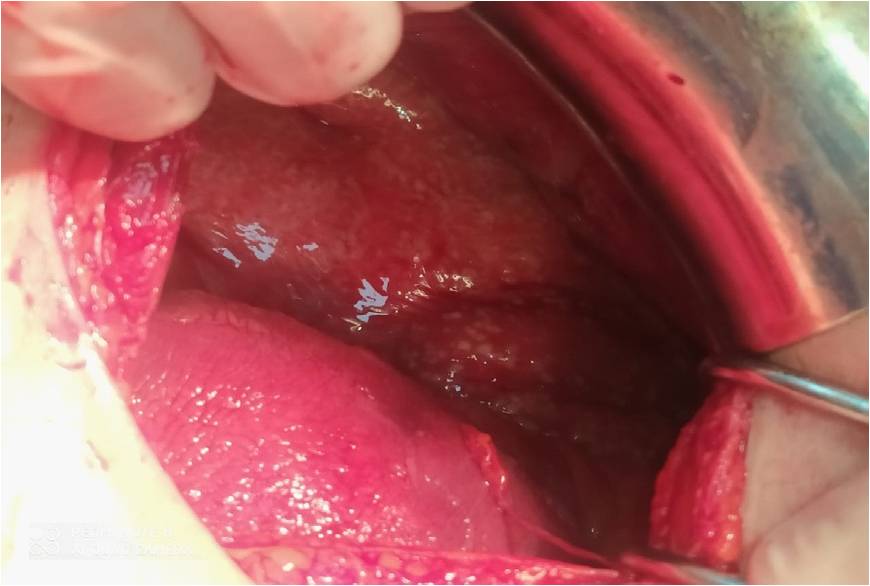
Many small white nodules were found with in the entire abdominal cavity, including the peritoneum, parietal and splanchnic (e.g. serosal surface of intestine, uterus, etc.), and greateromentum (Figures 1, 2). We performed a peritoneal and greateromentum biopsies.

Tuberculous peritonit is was highly suspected at the surgery, and thus, antituberculousis oniazid, rifampin, pyrazinamide, and ethambutol chemotherapy was promptly started. Histopathologic examination findings revealed numerous caseating chronic granulomatous inflammations caseo-follicular type in the biopsy and the identification of M. tuberculosis in ascites fluid.
The patient stayed at the intensive care unit for four days with good evolution and improvement of biological assessments and was shifted to the obstetrics unit ; she was declared discharged after five days.
The newborn was kept in neonatalogy unit for investigation and monitoring and put under preventive antituberculous treatment than got discharged with the mother.
Discussion
Tuberculous peritonitis is extremely rare in pregnancy, and the rate of TP among all forms of tuberculosis varies from 0.1% to 0.7% worldwide [1]. In literature there is a few cases of pregnancy with gestational ages between 20 and 24weeks of gestation [2,3]. In our case, gestational age was 34 weeks and vomiting started at 24 weeks of gestation.
Tuberculous peritonitis invades the entire abdominal cavity, it affects the parietal and visceral peritoneum, the omentum, the liver, the spleen, the intestinal tract and the pelvic cavity [4]. A possible mechanism to explain the pathogenesis of TP is the reactivation of latent tuberculous foci in the peritoneum or hematogenous spread from primary pulmonary tuberculosis [1]. However, the primary focus in the lungs is often healed completely, there by precluding its identification, despite careful examination of the patient [5].
The clinical symptoms of TP are non-specific, it is commonly associated with abdominal distension, pain, fever, chill, and weightloss. Our patient experienced most of those symptoms even so, we did not at any moment suspect the diagnosis of TP.
Gold standards for the diagnosis of TP are the identification of M. tuberculosis in ascites and peritoneal biopsy findings compatible with tuberculosis [1]. In fact, the positive ascites culture rate for M. tuberculosis is lessthan 50% [6] in our case the M. tuberculosis was identified in the ascites fluid.
Because of its clinical presentation (ascites, abdominal tenderness and abdominal masses), PT is often misdiagnosed as advanced ovarian cancer. Bacteriologic examination of the ascitic fluid is not always diagnostic [7] and clearly is not helpful when the patient is operated on an emergency basis.
Exploratory laparotomy or diagnostic laparoscopy. Bacteriologic examination of the biopsy specimen should be performed, because this could be positive for tuberculosis when histological examination is negative. This examination includes the identification of acid-fastbacilli (Ziehl—Neelsen staining positive), positive culture for M. tuberculosis and positive PCR for M. tuberculosis complex [8,9].
Conclusion
The diagnosis of TP in pregnancy is challenging, and therefore, it should be suspected when ascites and uncontrolled fever are present during pregnancy. Accurate diagnosis requires histopathologic examination and the isolation of M. tuberculosis from ascites and/or caseous necroticgranulomas from peritoneal biopsy nodules. The early diagnosis and treatment of TP in pregnancy are important to prevent adverse obstetric and neonatal morbidities.
References
- Sanai FM, Bzeizi KI. Systematic review: Tuberculous peritonitis: Presenting features, diagnostic strategies and treatment. Aliment Pharmacol Ther. 2005; 22: 685-700.
- Lahbabi M, Brini J, Massaoudi K. Tuberculous peritonitis in pregnancy: A case report. J Med Case Rep. 2014; 8: 3.
- Lee GS, Kim SJ, Park IY, Shin JC, Kim SP, et al. Tuberculous peritonitis in pregnancy. J Obstet Gynaecol Res. 2005; 31: 436-438.
- Koc S, Beydilli G, Tulunay G, Ocalan R, Boran N, Ozgul N, et al. Peritoneal tuberculosis mimicking advanced ovarian cancer: Are trospective review of 22 cases. Gynecol Oncol. 2006; 103: 565- 569.
- Marshall JB. Tuberculosis of the gastrointestinal track and peritoneum. Am J Gastroenterol. 1993; 88: 989-999.
- Avcu G, Sensoy G, Karli A, Caltepe G, Sullu Y, Belet N, et al. A case of tuberculous peritonitis in childhood. J Infect Public Health. 2015; 8: 369-372.
- Vardareli E, Kebapci M, Saricam T, Pasaoglu O, Ac¸ikalin, et al. M. Tuberculous peritonitis of the wet ascitic type: Clinical features and diagnostic value of image-guided peritoneal biopsy. Dig Liver Dis. 2004; 36: 199-204.
- Barutcu O, Erel HE, Saydili E, Yildirim T, Torun D. Abdomin opelvic tuberculosis simulating disseminated ovarian carcinoma with elevated CA125 level. Report of two cases. Abdom Imaging. 2002; 27: 465-470.
- Piura B, Rabinovich A, Leron E, Yanai-Inbar I, Mazor M. Peritoneal tuberculosis an uncommon disease that may deceive the gynecologist. Eur J Obstet Gynecol Reprod Biol. 2003;110: 230- 234.