
Journal of Clinical Images and Medical Case Reports
ISSN 2766-7820
Case Report - Open Access, Volume 2
Moth eaten alopecia as a manifestation of group of skin diseases: Reporting a series of 100 cases
Khalifa E Sharquie1; Ahmed Abdul-Aziz Ahmed2*
1 Department of Dermatology, College of Medicine, University of Baghdad, Medical City Teaching Hospital, Iraq.
2 Department of Dermatology, College of Medicine, Tikrit University, Iraq.
*Corresponding Author: Ahmed Abdul-Aziz Ahmed
Department of Dermatology, College of Medicine,
University of Baghdad, Medical City Teaching Hospital,
Iraq.
Email: ahmedabdul-aziz@tu.edu.iq
Received : Nov 11, 2021
Accepted : Dec 17, 2021
Published : Dec 24, 2021
Archived : www.jcimcr.org
Copyright : © Ahmed AA (2021).
Abstract
Background: The moth-eaten alopecia is a characteristic clinical pattern usually of multiple small ill-defined patches of hair loss that was originally used to describe alopecia in syphilis but it is not well reported in other skin diseases.
Objective: To record all patients that present with moth eaten alopecia and to do full clinical evaluation and classification aiming to find skin diseases that initially or during the course of the disease present with this pattern of hair loss.
Patients and methods: This is a descriptive observational study of a series of 100 patients with moth-eaten alopecia. The data were collected in Baghdad city from 2014 to 2021 with an age range between 8-45 years involving 72 males and 28 females. Full laboratory evaluation including scraping for fungal infection and skin biopsy were performed when its indicated and justified.
Results: The results of this study showed that lichen planopilaris, was recognized in 32(32%) patients, their ranged from 20-45 years with a mean of 25, while males consisted 31(96.8%) and females 1(3.2%). Discoid lupus erythematous was identified in 26(26%) patients, their ages ranged from 18-48 years with a mean 30 years including 7(26.9%) males and 19(73.1%). Females. Alopecia areata was recognized in 11(11%) patients, their ages ranged from 8-25 years with a mean 17 years, among them 6 males (54.5%) and 5 females (45.5%). Chronic folliculitis presented in 9(9%) male patients, their ages ranged from 20-30 years with a mean 25 years. Striae distensea was seen and identified in 9 patients their ages ranged from 3-10 years with a mean 7 years, among them males 8(89%) and females 1(11%). Scalp morphea was detected in 7(7%) patients, their ages ranged from 5-34 years with a mean of 29 years among them 5(71.5%) males and 2 (28.5%) females. Tinea capitis was seen in 4(4%) male patients, their age ranged from 3-5 years with a mean age of 4 years. While Juicy Psoriasis was encountered in two male patients (2%). Their ages ranged from 7 and 15 years with a mean 11 years.
Conclusion: This is the first study describing and evaluating moth eaten alopecia as a characteristic and unique pattern of hair loss for many dermatosis. lichen planopilaris, discoid lupus erythematous, chronic folliculitis, striae distensae like were among the commonest diseases that cause this type of hair loss. This work will provide an important differential and diagnostic tool for clinician in order to establish early diagnosis and treatment, thus preventing the progress of the disease and scarring.
Citation: Sharquie KE, Ahmed AA. Moth eaten alopecia as a manifestation of group of skin diseases: Reporting a series of 100 cases. J Clin Images Med Case Rep. 2021; 2(6): 1510.
Introduction
Syphilitic Alopecia (SA) is rare, occurring in 2.9-7% of syphilic patients [1]. As first described by McCarthy in 1940, syphilitic alopecia can be categorized into symptomatic alopecia and essential alopecia. The symptomatic presented as patchy alopecia associated with papulosequamous skin lesions [2]. The essential kind can be divided into moth-eaten alopecia, diffuse telogen effluvium like alopecia and mixed pattern. The motheaten alopecia is a distinguishing clinical fashion of multiple small ill-defined patches of hair loss that was originally used to describe the most frequent pattern of alopecia in syphilic patients [2,3]. It mainly occur on the occipito-parietal region due to rich blood supply nevertheless can also occur in other hairy areas. Immunohistochemical studies uncovered the presence of spirochete in the hair follicle, accordingly confirming that hair loss is secondary to immunological reaction to the presence of spirochetes [4]. In addition, the patient routinely presents with systemic manifestations of secondary syphilis like fever, malaise, generalized lymphadenopathy and nonpruritic copper-colored papulosquamous rash. On histopathology, moth-eaten alopecia is characterized by perifollicular lymphohistocytic infiltration with prominent plasma cells. Rarely, primary and tertiary syphilis may cause alopecia but only when occurs on the scalp5. Treatment of SA with penicillin resulted in complete regrowth of the hair [3-5].
After considerable review of the medical literatures, there were no established studies to describe this pattern of hair loss in dermatosis other than syphilis, Apart from a study by Sharquie et al in 2013 [6] described extensively moth-eaten alopecia in lichen planopilaris. Since syphilis is now almost rare and may have been finished in many countries including Iraq, this study aims to collect cases with moth-eaten pattern of hair loss and to do full evaluation of these disorders.
Patients and methods
This is a descriptive observational study of a series of 100 patients with moth-eaten alopecia pattern. The data were collected in Baghdad city from 2014 to 2021 with an age range between 8-45 years involving 72 males and 28 females. Full laboratory evaluation including scraping for fungal infection and skin biopsy were performed in indicated cases. Data were presented in numbers and percentages. Informed consent was obtained for all patients involved in this study.
Results
The present study included a series of 100 patients with typical moth eaten alopecia, their ages ranged from 3-48 years with a mean age 45 years among them 72 males (72%) and 28 females (28%).
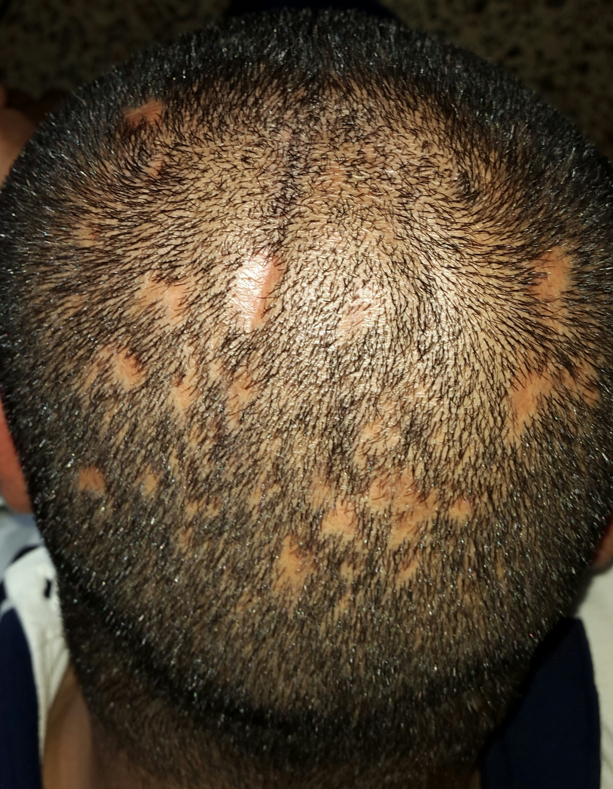
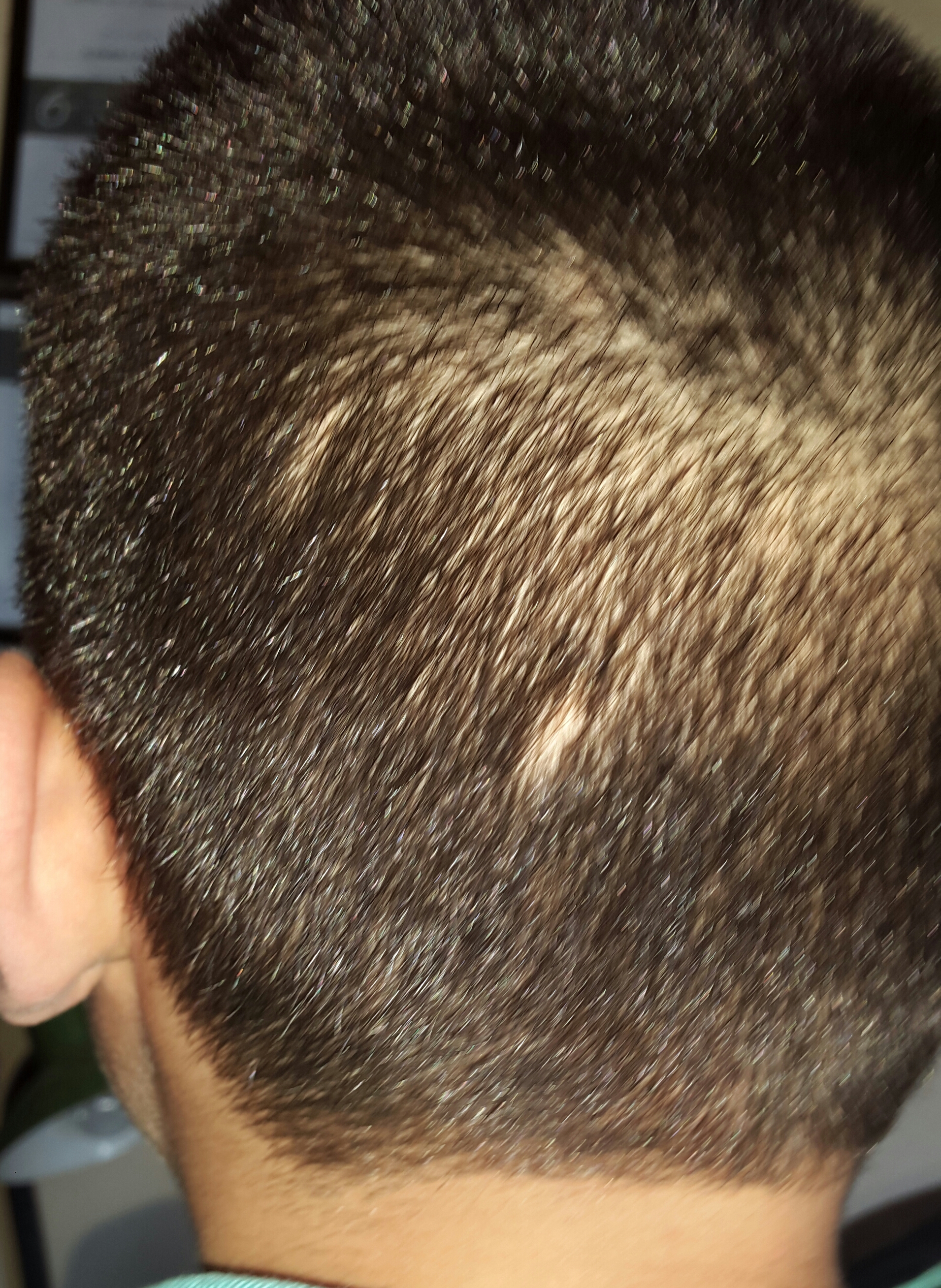
This study showed the following skin diseases: Lichen planopilaris was seen and recognized in 32(32%) patients, their ranged from 20-45 years with a mean of 25 year, among them 31(96.8%) males and 1(3.2%) female. Clinically the disease started as small follicular lesions that after a while coalesced together to form distinct and unique moth eaten alopecia pattern of hair loss and then extend to involve large areas leaving characteristic pigmented scarring with some islands of normal hair what was appearing as fire in field (Figure 1).
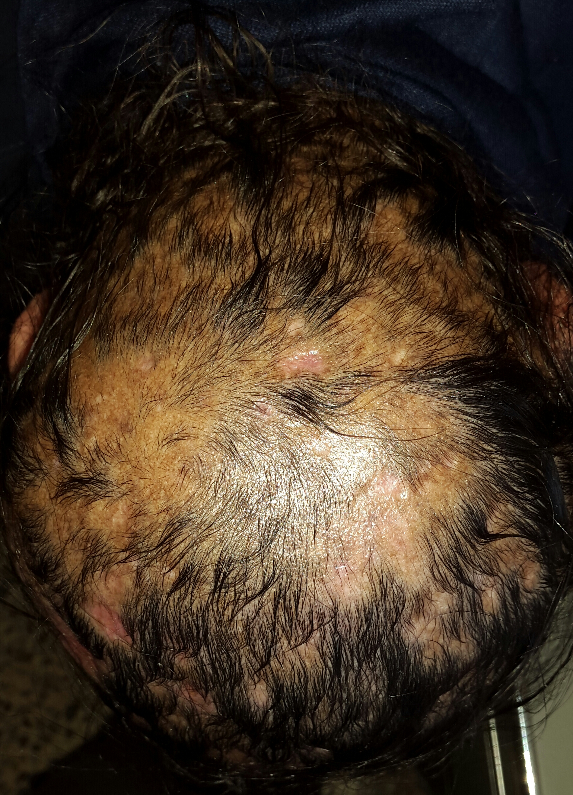
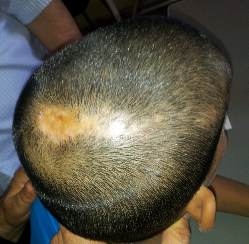
Discoid lupus erythematous was identified in 26 (26%) patients their ages ranged from 18-48 years with mean of age 30 years among them 7(26.9%) males and 19(73.1%) females. It was observed that the hair loss initially started as small irregular patches of moth eaten alopecia then overtime merged together to form atrophic erythematous or leukodermic patches (Figure 2).
Alopecia areata was detected in 11(11%) patients, their ages ranged from 8-25 years with a mean of age 17 years. There were 6(54.5%) males and 5(45.5%) females. These patients presented with moth eaten alopecia with completely normal scalp apart from exclamation mark hairs that regarded the hallmark sign in these cases (Figure 3).
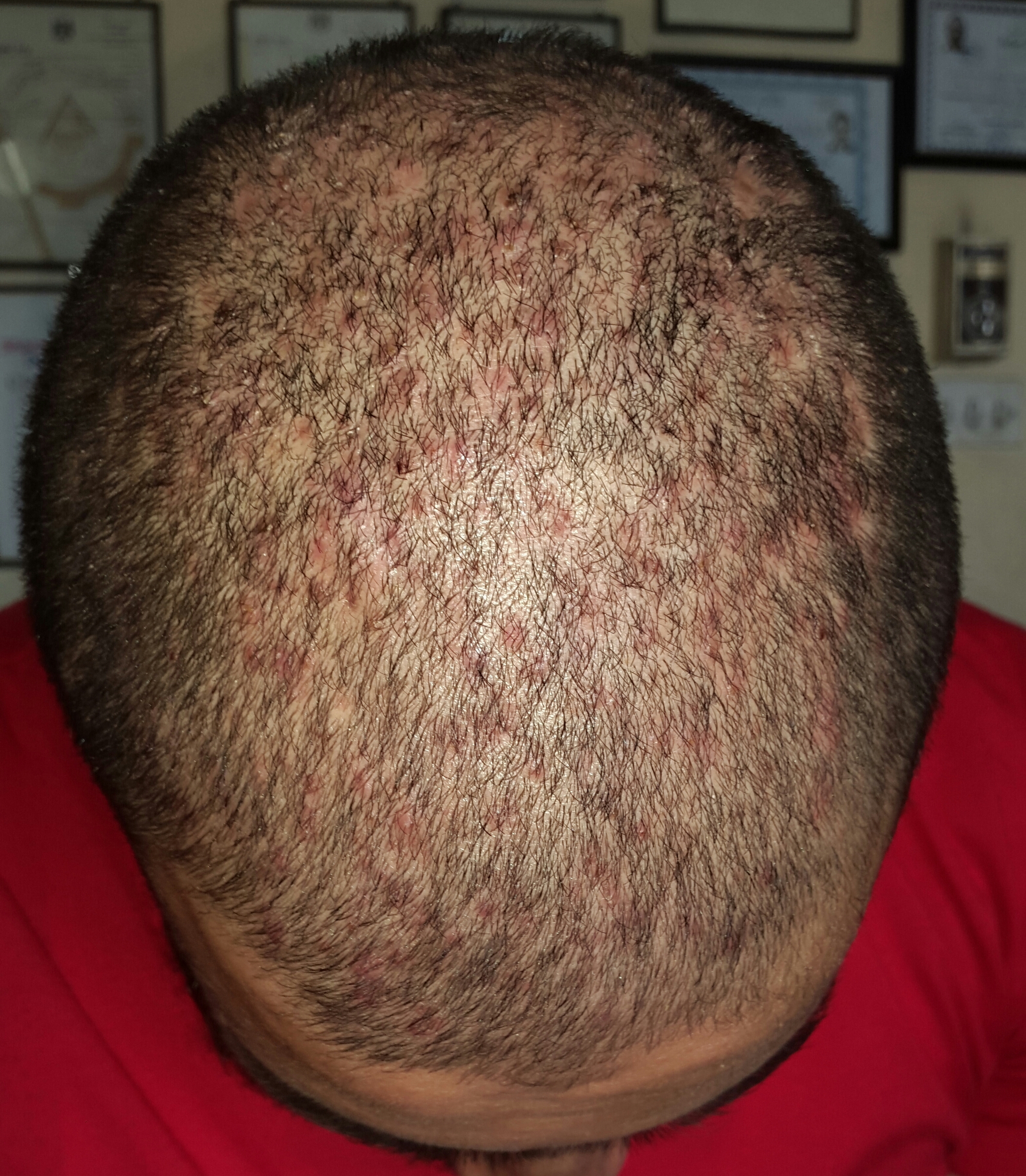
Chronic folliculitis was observed in 9(100%) male patients, their ages ranged from 20-30 years with a mean 25 years. The condition initially started as painful follicular papules and pustules that after healing left moth eaten alopecia and this inflammatory process could be repeated over time leaving extensive hair loss which was usually none scarring (Figure 4).
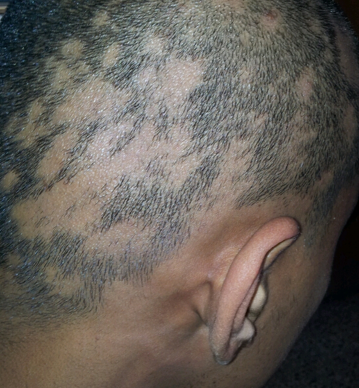
Striae distensea was identified in 9(9%) patients, their ages ranged from 3-10 years with a mean of 7 years, among them 8 (89%) males and 1(11%) females. All patients were treated as alopecia areata but no recovery. Clinically presented as single or multiple linear hair loss with 1-6 lesions, resembling atrophic scars, slightly reddish in color which was similar to striae distensae (atrophicans). They were completely free of hair and without broken and or exclamation mark hairs at the margin of the linear patches. The size of the patches was varied from 1-3 cm in length and the width usually ranged from 0.5-1 cm. Full examination of the whole skin showed no striae distensae in all patients. On careful questioning, their parents denied any history of obvious trauma (Figure 5).
Scalp morphea was observed in 7 patients (7%), their ages ranged from 5-34 years with a mean of 29 years among them 5 (71.5%) males and 2 (28.5%) females. Clinically the condition started as slightly erythematous patches and then gradually and slowly became depressed and atrophic linear scars with a patchy hair loss in small areas simulating moth eaten alopecia in early course of the disease. It was worth noting that in all recorded cases were linear morphoea in a form of en coup de sabre (Figure 6).
Tinea capitis was seen in 4(4%) male patients, their age ranged from 3-5 years with a mean age of 4 years. Clinically, we noticed that patients with diffuse scaly scalp that involved part or most of the scalp and then overtime there was a partial hair loss in small areas in picture of moth eaten alopecia (Figure 7). Juicy psoriasis was encountered in two male patients (2%) their ages 7 and 15 years with a mean of 11 years (Figure 8).
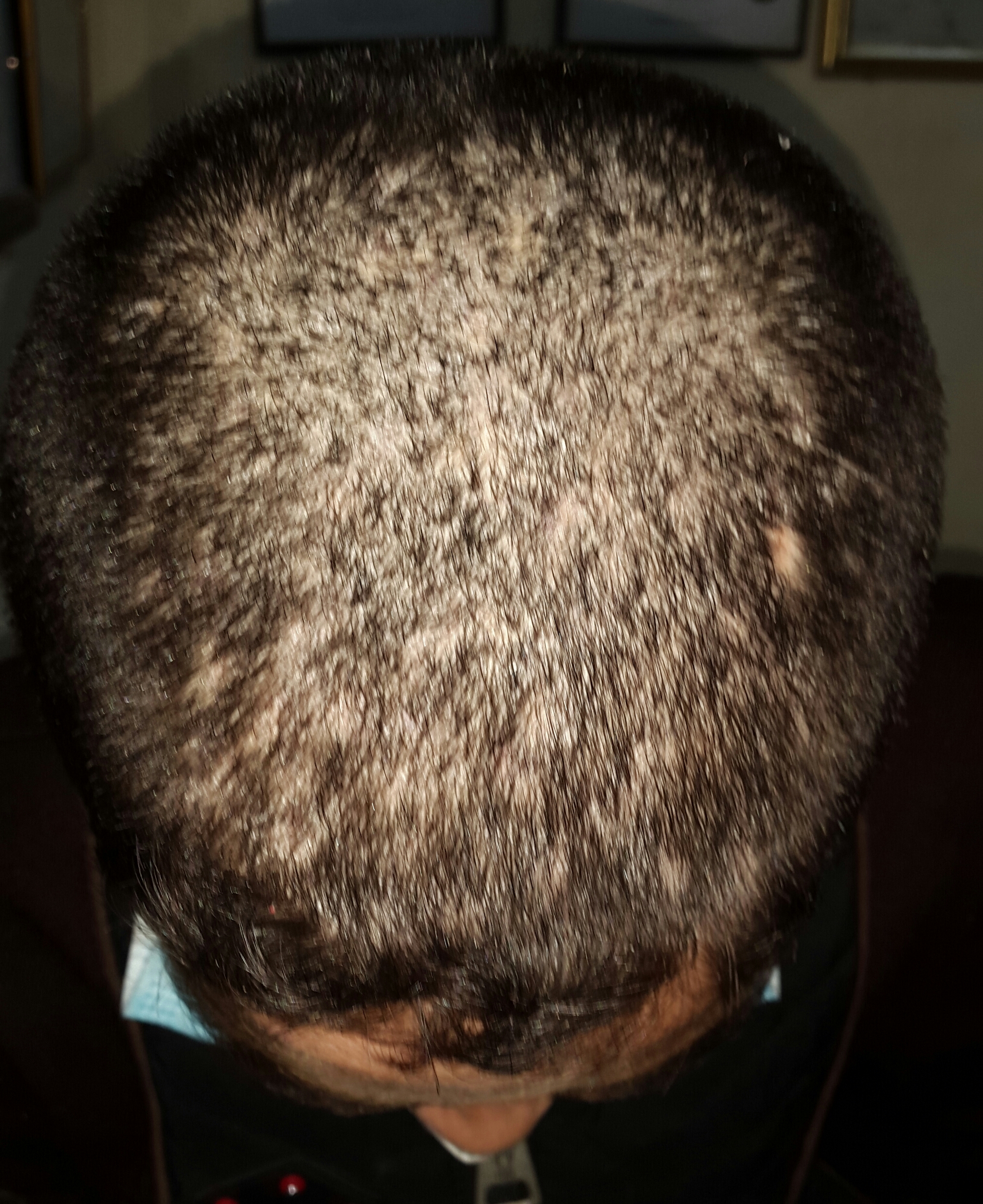
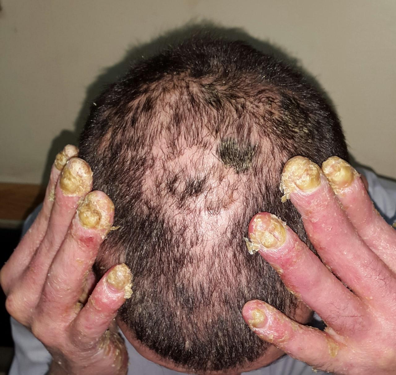
Table 1: Frequency of moth-eaten alopecia among all patients.
Age (years) |
Mean age (years) |
Males |
Females |
Total |
||
No |
No |
No |
% |
|||
lichen planopilaris |
20-45 |
25 |
31 |
1 |
32 |
32 |
Discoid lupus erythematosus |
18-48 |
30 |
7 |
19 |
26 |
26 |
Alopecia areata |
8-25 |
17 |
6 |
5 |
11 |
11 |
Chronic folliculitis |
20-30 |
25 |
9 |
0 |
9 |
9 |
Striae distansae |
3-10 |
7 |
8 |
1 |
9 |
9 |
Scalp morphea |
5-34 |
29 |
5 |
2 |
7 |
7 |
Tinea capitis |
3-5 |
2 |
4 |
0 |
4 |
4 |
Juicy psoriasis |
7- 15 |
11 |
2 |
0 |
2 |
2 |
Total |
|
|
72 |
28 |
100 |
100 |
Discussion
Moth-eaten alopecia is a common and extensively faced problem in dermatological practice in which many diseases sharing this feature. To the best of our knowledge, the current work represent the first effort in medical literatures to trace and describe this group of dermatosis with moth-eaten alopecia pattern. Since syphilis is now extremely rare in Iraq and in many other countries, it is essential and of potential clinical importance to recognize the other hair loss diseases that has a moth-eaten pattern and to provide early diagnosis which is essential for the initiation of an early and effective therapy to save the follicular apparatus in certain hair loss disorders in the scalp which may start as moth eaten alopecia, thus preventing the progression of the disease and further scarring.
Regarding, Lichen planopilaris (LPP) classically presents as follicular keratotic plugs and/or perifollicular scaling along with perifollicular erythema, with subsequent hair loss resulting in patchy alopecic areas [7,8]. Histologically, the active and early lesion show degenerative basal keratinocytes and destruction of the basal layer in half cases and a band-like subepidermal lymphocytic infiltrate, “hugging” the upper hair follicle (isthmus and infundibulum), with no involvement of the deeper portion of the follicle. While in late-stage lesions, inflammation can be minimal or absent, with no distinctive lichenoid changes, fibrous tracts replaced the destroyed hair follicles. And reduction/loss of sebaceous glands and arrector pili muscles, concentric perifollicular fibrosis, and irreversible destruction of the follicle with perifollicular hyalinization in both upper/lower dermis and follicular tract. Other specific histological features include mucinous perifollicular fibroplasia in the upper dermis, the absence of interfollicular mucin, and a superficial perifollicular wedge-shaped scarring [7-9]. Sharquie et al in 2013 described 67 patients with moth-eaten lichen planopilaris [6]. The age range from 20 to 65 (mean 36.7+5) years and the male to female ratio was 1.4:1 (39 males vs 28 females). The author described the clinical pattern of alopecia as moth eaten alopecia. The present case series study revealed that lichen planopilaris is the most common cause of moth-eaten alopecia accounting for 32% (n=32) of patients with age range is 20-45 years. So the current study is in complete agreement with Sharquie's previous study.
Discoid Lupus Erythematosus (DLE) of the scalp is a typical form of scarring alopecia, characterized by well-defined erythematous plaques with scales, atrophy, follicular plugging, central hypopig-mentation, and peripheral hyperpigmentation. Lesions develop progressively from non-cicatricial alopecia to a late scarring phase.
Histopatholgically, hyperkeratosis with follicular plugging thinning and flattening of the stratum malpighii, hydropic degeneration of basal cells, dyskeratosis, and a predominantly lymphoplasmacytic infiltrate arranged along the dermalepidermal junction, around hair follicles and eccrine glands with interstitial mucin deposition, and a possible slight extension of the inflammatory infiltrate to the Subcutaneous [11].
DLE should be differentiated from LPP both clinically and histologically.
In which Clinically early lesions of DLE are scaly erythematous with central activity of the lesions and scarring develop late in the course of disease, While early lesions of LPP are pigmented follicular with peripheral activity of the lesions and scarring develop early in the course of disease.
Histopatholgically, regarding DLE characterized by vascular interface dermatitis, normal granular layer with inflammation of the entire follicle mostly around the isthmus and peri eccrine inflammation with thick basement membrane with involvement of the interfollicular epidermis and superficial and deep perivascular infiltrate and panniculitis. on the other hand LPP characterized by vascular interface dermatitis, wedge hypegranulosis with inflammation of the upper half of the follicle mostly around the infundibulum and sparing the eccrine gland with normal basement membrane with sparing the interfollicular epidermis and superficial perivascular infiltrate and no panniculitis.
Alopecia areata present as single or multiple well defined patches of complete non-cicatricial hair loss. It can also involve the entire scalp (alopecia totalis) or the entire body (Alopecia universalis). A moth-eaten alopecia like pattern was also described by the current work. The characteristic exclamation marks can be observed in most patients. Histologic examination reveals swarm of bee pattern of peribulbar lymphocytic infiltration with characteristic eosinophils and miniaturization of hair follicles[5]. Although moth-eaten in syphilis might mimic alopecia areata, but those of syphilis are characterized by prominent plasma cell infiltration without eosinophils or hair follicle miniaturization [12]. Moth eaten alopecia in alopecia areata was not reported in medical literature.
Chronic folliculitis is common problem especially among adult males where it starts initially as follicular papules and pustules that after recovery ends with moth eaten alopecia and this picture of hair loss might confused with other causes of patchy hair loss. This new pattern of hair loss was not recognized and published before the present study [13].
Striae distensae-like lesions are a new entity, which seems to be common among children and often confused with untreated cases of alopecia areata which was first described and illustrated by Sharquie et al [14]. There is no history of trauma to scalp but we can speculate that, occasionally, rupture dermal connective tissue caused by blunt trauma but not injuring epidermis so no bleeding to be seen [14]. It is worth mentioning that this new entity might simulate, both clinically and histopathologically, lichen sclerosis but this proposal might need further exploration. This condition may presented with moth eaten alopecia pattern and should be included in the differential diagnosis of patchy hair loss with moth eaten alopecia especially in children. The parents should be reassured of the cause of hair loss and no treatment is required.
Although scalp morphea is reported in the literatures, but it's presentation with moth-eaten alopecia was never mentioned. It is well known that scalp morphea induces alopecia either in linear (en cop de sabre) or circumscribed pattern [15]. The present study reports the unique pattern of mouth eaten alopecia induced by localized scleroderma.
Various clinical patterns are described with tinea capitis: 1) Black dot after broken off hair shafts close to the scalp, the left-over hair follicles are seen as prominent black dots. 2) Gray patch Prominent scales in patchy alopecia with or without erythema. 3) Diffuse pustular Scattered pustules with scale, alopecia, and lymphadenopathy. 4) Diffuse scale Large areas of scaling of scalp with or without erythema. 5) Kerion Well-defined bald area with red, boggy, pus-filled tender plaque, and lymphadenopathy [18].
In the current study we reported for a first time a cases of tinea capitis presented clinically with hair loss in a moth-eaten alopecia pattern and confirming the diagnosis with Microscopic examination for hyphae and culture.
Finally, also we have reported two cases of Juicy psoriasis.
Conclusion
Mouth-eaten alopecia of the scalp is a unique and distinctive model of hair loss that's currently common in clinical presentation due to variety of hair loss disorders but lichen planopilaris, discoid lupus erythematous, chronic folliculitis, striae distensae like are among the commonest diseases that cause this type of hair loss. To the best of our knowledge, this is the first study reporting and evaluating this characteristic pattern of hair loss. The current work will facilitate early diagnosis and treatment, thus preventing the progress of the disease and scarring.
References
- Doche I, Hordinsky MK, Valente NY, Romiti R, Tosti A. Syphilitic alopecia: case reports and trichoscopic findings. Skin appendage disorders. 2017; 3: 222-4.
- McCarthy L. Diagnosis and treatment of diseases of the hair. St. Louis: CV Mosby; 1940: 537-48.
- Cuozzo DW, Benson PM, Sperling LC, Skelton HG. Essential syphilitic alopecia revisited. J Am Acad Dermatol. 1995; 32: 840-3.
- Expósito V, Iglesias M, Solà J, Dilme E, Umbert P. Alopecia sifilítica esencial: forma única de presentación de un secundarismo luético. Piel. 2011; 26: 305-7.
- Mindel A, Tovey SJ, Timmins DJ, Williams P. Primary and secondary syphilis, 20 years’ experience. 2. Clinical features. Genitourin Med. 1989; 65: 1-3.
- Sharquie KE, Noaimi AA, Hameed AF. Lichen Planopilaris is a Common Scarring Alopecia among Iraqi Population. Journal of Cosmetics, Dermatological Sciences and Applications. 2013; 3(3): 35-9.
- Lyakhovitsky A, Amichai B, Sizopoulou C, Barzilai A. A case series of 46 patients with lichen planopilaris: demographics, clinical evaluation, and treatment experience. J Dermatolog Treat. 2015; 26(3): 275-279.
- Assouly P, Reigagne P. Lichen planopilaris: update on diagnosis and treatment. Semin Cutan Med Surg. 2009; 28(1): 3-10.
- Rácz E, Gho C, Moorman PW, Noordhoek Hegt V, Neumann HA. Treatment of frontal fibrosing alopecia and lichen planopilaris: a systematic review. J Eur Acad Dermatol Venereol. 2013; 27(12): 1461-1470.
- Garbelini-Lima C, de Almeida GE, Gabdelha SQ, de Souza AC, de Souza ML, Figueiras VV. Discoid Lupus Erythematosus of the Scalp in a Patient with Systemic Lupus Erythematosus: A Case Report with Complete Hair Regrowth. Journal of the Portuguese Society of Dermatology and Venereology. 2021; 79(2): 155-8.
- Harry Winfield, Christine Jaworsky. Connective Tissue Diseases, David E. Elder, Lever’s Histopathology of the Skin, 11th edition, 2015.
- Gilhar A, Etzioni A, Paus R. Alopecia areata. New England Journal of Medicine. 2012; 366(16): 1515-25.
- Zulian F. Systemic manifestations in localized scleroderma. Curr Rheumatol Rep. 2004; 6(6): 417-24.
- Sharquie KE, Al-Waiz MM, Al-Nuaimy AA. Striae distensae-like lesions. A cause of scarring alopecia among children. Saudi medical journal. 2002; 23(12): 1489-91.
- Florez-Pollack S, Kunzler E, Jacobe HT. Morphea: Current concepts. Clinics in dermatology. 2018; 36(4): 475-86.
- Bennassar A, Grimalt R. Management of tinea capitis in childhood. Clinical, cosmetic and investigational dermatology: CCID. 2010; 3: 89
- Sah DE, Koo J, Price VH. Trichotillomania. Dermatologic therapy. 2008; 21(1): 13-21.
- Chitkara S, Kaur R, Mehra B, Chitkara AJ. Fungal Infections in Children: A Simplified Approach. Pediatric Infectious Disease. 2021; 3(1): 27.