
Journal of Clinical Images and Medical Case Reports
ISSN 2766-7820
Case Report - Open Access, Volume 2
A case of unruptured tectal located AVM presenting with obstructive hydrocephalus
Ozlem Kadirhan, MD1*; Sonay Aydin, MD1; Erdal Karavas, MD1; Mecit Kantarci, MD1,2
1 Department of Radiology, Faculty of Medicine, Erzincan University, Erzincan, Turkey
2 Department of Radiology, Faculty of Medicine, Ataturk University, Erzurum, Turkey.
*Corresponding Author: Ozlem Kadirhan
Department of Radiology, Faculty of Medicine,
Erzincan University, Erzincan, Turkey.
Email: ozlemkkadirhan@gmail.com
Received : Dec 11, 2021
Accepted : Jan 12, 2022
Published : Jan 19, 2022
Archived : www.jcimcr.org
Copyright : © Kadirhan O (2022).
Keywords: arteriovenous malformation; obstructive hydrocephalus; tectum mesencephali.
Abbreviations: AVM: arteriovenous malformations; CT: computed tomography; CTA: computed tomography angiography; MRI: magnetic resonance imaging.
Citation: Kadirhan O, Aydin S, Karavas E, Kantarci M. A case of unruptured tectal located AVM presenting with obstructive hydrocephalus. J Clin Images Med Case Rep. 2022; 3(1): 1583.
Clinical image description
We present a case of arteriovenous malformation with aneurysmatic dilatation of the drainage vein, presenting with obstructive hydrocephalus located in the tectum.
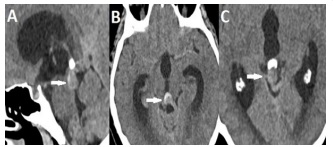
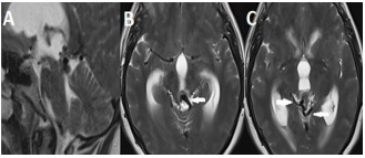
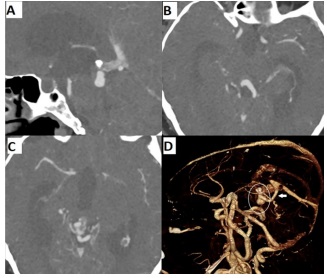
A 59-year-old female patient was taken to the emergency department after complaining for 8 days of increasingly worsening headache, gait disturbance, sleepiness, blurred vision, and double vision, as well as gush vomiting. The patient indicated that he had been experiencing from intermittent headaches for almost a year. On examination, there was an ataxic walk, bilateral papilledema, and slight cognitive impairment. CT, MRI and CTA imaging tests were used for the diagnosis, respectively (Figures 1-3). CT scans of the brain revealed triventricular hydrocephalus and a dense space occupying lesion thought to produce aqueductal stenosis in the tectal region of the mesencephalon (Figure 1). Following that, the patient had brain MRI and CTA imaging. This dense lesion in the tectum was found to be a vascular structure connected to the adjacent arteriovenous capillary network and the vein of galena (Figure 2,3). In the differential diagnosis, arteriovenous malformation with aneurysmatic dilatation in the drainage vein was considered primarily.
AVMs are a rare vascular disorder of the central nervous system that most commonly presents as a congenital intracranial shunt through the nidus between the arterial and venous systems that lacks a capillary bed. A large retrospective study has shown a lower incidence of AVMs located in the brainstem (4%) [1]. While AVM is most commonly manifested as bleeding, its relationship with hydrocephalus is uncommon, and this link is most commonly caused by a ruptured AVM [2]. The case we presented was an AVM located in the mesencephalon, which caused hydrocephalus without rupture.
Declarations
Ethics approval and consent to participate: All procedures performed in studies involving human participants were in accordance with the ethical standards of the institutional and/ or national research committee and with the 1964 Helsinki declaration and its later amendments or comparable ethical standards.
Consent for publication: All appropriate consent forms were obtained from the patient. On the form, he gave consent for patient pictures and other clinical information to be reported in the journal. The patient understood that his name and initials would not be published and that every effort would be made to conceal his identity.
Competing interests: The authors declare that they have no competing interests.
Funding: No funding have been received for the study.
References
- Lawton MT, Rutledge WC, Kim H, Stapf C, Whitehead KJ, Li DY et al. Brain arteriovenous malformations. Nat Rev Dis Primers. 2015; 1(1): 1-20.
- Mindea SA, Yang BP, Batjer HHJNf. Unruptured arteriovenous malformation in a patient presenting with obstructive hydrocephalus: Case report and review of the literature. Neurosurg Focus. 2007; 22(4): 1-4.