
Journal of Clinical Images and Medical Case Reports
ISSN 2766-7820
Clinical Image - Open Access, Volume 3
Intraosseous hydatid cyst
Amarkak Abdillahi Wais*; Daoud Mohamed Ali; Soufiane Rostoum; Assad El Bakkari; Laila Jrondi; Fatima-Zahra Laamrani
Department of Radiology, CHU Ibn-sina, Mohamed V University, Rabat, Morocco.
*Corresponding Author: Amarkak Abdillahi Wais
Department of Radiology, Hospital of Specialty, CHU
Ibn-Sina, Mohamed V University, Rabat Morocco.
Email: amarkak.a.wais@gmail.com
Received : Jan 15, 2022
Accepted : Feb 21, 2022
Published : Feb 28, 2022
Archived : www.jcimcr.org
Copyright : © Wais AA (2022).
Citation: Wais A, Ali WD, Rostoum S, Bakkari AE, Jrondi L, et al. Intraosseous hydatid cyst. J Clin Images Med Case Rep. 2022; 3(2): 1695.
Description
Hydatidosis is a parasitic disease caused by Echinococcus granulosus or echinococcal tænia, preferentially localized in the lung (20 to 30%) and in the liver (60 to 70%). Intraosseous localization is extremely rare.
She did not complain on any difficulties swallowing or breathing. She has a background of mild Asthma for which she uses inhalers but otherwise is generally well in herself. She is a non-smoker and is fully vaccinated.
Imaging features takes a very important place for the diagnosis and therapeutic management such as ultrasound, CT and MRI.
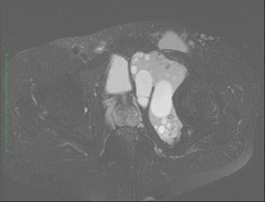
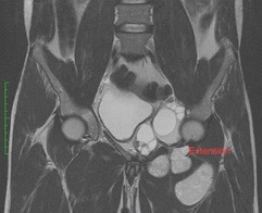
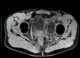
MRI shows a large, well-limited and multiloculated cystic mass in cotyloid cavity, with daughter vesicles in hypo-intensity T1-weighted, hyperintensity T2-weighted with low diffusion coefficient and no contrast enhancement after injection of gadolinium. This mass causes a breach of cortex and extension to the adjacent soft tissue.
References
- Ladjouze Rezig A. Hydatidose osseuse. Rev Rhum 2002; 69: 835- 84.
- Bel Hadj Youssef D et al. Kyste hydatique primitif intraosseux: A propos de deux cas. Rev Med Interne. 2007; 28: 255-258.
- Hasan Onur Arik Primary intraosseous hydatid cyst of femur: Iran Red Crescent Med J. 2015; 17: e21070.