
Journal of Clinical Images and Medical Case Reports
ISSN 2766-7820
Case Series - Open Access, Volume 3
Rare benign tumors of the nipple: Report of 2 cases
Hamideh Mohammadzadeh1; Naser Tayyebi Meibodi2; Ehsan Noori3; Maliheh Amani1*
1 Department of Dermatology, School of Medicine, Gonabad University of Medical Sciences, Gonabad, Iran.
2 Cutaneous Leishmaniasis Research Center, Mashhad University of Medical Sciences, Mashhad, Iran.
3 Department of Anesthesiology, Imam Reza Hospital, Mashhad University of Medical Sciences, Mashhad, Iran
*Corresponding Author: Maliheh Amani
Department of Dermatology, School of Medicine,
Gonabad University of Medical Sciences, Gonabad,
Iran.
Email: ml.amani@yahoo.com
Received : Feb 26, 2022
Accepted : Mar 25, 2022
Published : Apr 01, 2022
Archived : www.jcimcr.org
Copyright : © Amani M (2022).
Abstract
Fibroepithelial of polyp and epidermal inclusion cyst are common benign skin lesions but they arise rarely on nipple. This paper reports rare manifestations of these common lesions. The first patient manifested with huge polypoid mass on nipple that microscopic examination proved fibroepithelial of polyp. The second patient had a smooth papule on her nipple that was diagnosed as Epidermal inclusion cyst.
Citation: Mohammadzadeh H, Meibodi NT, Noori E, Amani M. Rare benign tumors of the nipple: Report of 2 cases. J Clin Images Med Case Rep. 2022; 3(4): 1771.
Introduction
Nipple is a central part of breast which is drain path of milk. Differential diagnoses of nipple lesions are various, some lesions rarely presented on it [1]. Fibroepithelial of polyp and epidermal inclusion cyst are benign tumors that rarely appear on nipple. Fibroepithelial of polyp (skin tag) is a type of mesenchymal tumor which is benign and asymptomatic and rarely exceeds 2 cm. This tumor develops more on some areas such as face, neck, axilla and groin [2]. Epidermal inclusion cyst is the most common cutaneous cyst that a rise spontaneously or post trauma implantation. Although it is a common lesion, Nipple is an unusual site for it [3]. Here in we report 2 benign lesions (a giant fibroepithelial of polyp and an epidermal inclusion cyst) that are rarely developed on nipple.
Case presentations
Case 1
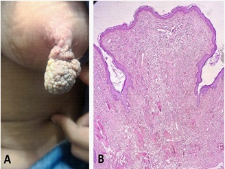
A 24-year-old woman presented to dermatology office with a mass on her left nipple which was pedunculated with lobulated surface (Figure 1A). She didn’t complain of pain, bleeding or discharge. It had appeared since age of 13 that had slow growth but had enlarged rapidly during last year. It was a size 22 X 22 X 20 mm. It didn’t have any rapid growth or involution during the pregnancy and breast feeding. On Physical examination, both of the breasts and the other nipple were normal and we didn’t touch any mass. We didn’t detect any enlargement of axillary lymph nodes. Medical history was normal. Familial history of breast cancer was negative. She had taken no medications. Breast imaging was otherwise unremarkable. Microscopic examination by H&E staining showed a polypoid tumor with flattened epidermis overlying fibrovascular tissue in the dermis. The vessels were Dilated and thin-walled. These microscopic features proved the diagnosis of fibroepithelial of polyp (Figure 1B). We excised the tumor completely under local anesthesia while the nipple was spared. She recursed our clinic after 9 month and recurrence was not observed.
Case 2
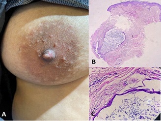
A 33-year-old woman presented with a papule on her right nipple. It had been appeared for 2 months. She didn’t complain any pain and discharge. On physical examination, it was immobile with smooth surface and its size was 5 X 4 X 3 mm (Figure 2A). Both of the breasts and the other nipple were normal and we didn’t touch any mass as well. She had breastfed two months prior to appearance the lesion. She noted no Familial history of breast cancer. Medical history was normal. She had taken no medications. Breast sonography was otherwise normal. We obtained a skin biopsy. Microscopic examination by H&E staining showed a cystic structure that lined by squamous epithelium and is filled with keratin with no connection to hair follicle (Figure 2B,C). These histologic features made diagnosis of epidermal inclusion cyst. The cyst was excised completely.
Discussion
Although benign tumors of nipple are much more frequent than malignant ones, any Nipple lesions specially in women might cause concern, because there is a possible connection between nipple lesions and breast malignancy. So definite diagnosis of nipple lesions is challenging and critical. Some manifestations such as epidermal changes, abnormal discharge, bleeding, pain and nipple inversion are more common in malignant tumor, high age and rapid enlarged lesions are risk factors as well [2,4]. We evaluated the patients completely and both of our patients had no clues in favor of malignant lesions on history, physical examination and imaging. Fibroepithelial of polyp fairly develops on nipple and rarely exceeds 2 cm, so larger lesions are called giant skin tag. Ever since just a few giant fibroepithelial polyps of nipple have been reported in literatures [2,5,6]. Our patient complained a huge mass on nipple which had enlarged rapidly during last year. This case presented with unusual size on uncommon site of fibroepithelial of polyp. Epidermal inclusion cyst originates from infundibulum of hair follicle so they present on hairy body areas. Nipple is a hair-free area and unusual site for arising this cyst [7]. How does it develop on nipple? Some authors have theorized that epidermal inclusion cysts on nipple may be result of metaplasia of ductal epithelial of the breast (columnar epithelium to stratified squamous epithelium) [8]. In microscopic examination of our case, the cyst locates deeply near the ductal epithelial of the breast and there is no hair follicle for connection to it. To our knowledge, a few cases of epidermoid cysts on nipple have been reported and the most of them were post traumatic [3]. Our patient didn’t note any history of trauma, but she had breastfed two months prior to appearance the lesion. This is possible the breastfeeding might cause the trauma.
References
- Cai S, Wang H, Zhu Q, Li J, Sun Q, et al. Clinical and sonographic features of nipple lesions. Medicine. 2020; 99.
- Hameed OF, Maktoof ZA, Husien HF. Fibroepithelial polyp in a female right breast nipple-Case Report. Journal of Medical Research and Health Sciences. 2021; 4: 1377-1380.
- Jain S, Sarkar R, Garg V, Khurana N. Epidermal inclusion cyst or giant milium of the nipple. Indian Journal of Dermatology Venereology and Leprology. 2012; 78: 103.
- Spyropoulou GA, Pavlidis L, Trakatelli M, Athanasiou E, Pazarli E, et al. Rare benign tumours of the nipple. Journal of the European Academy of Dermatology and Venereology. 2015; 29: 7-13.
- Arora BK. A clinical case of large fibroepithelial polyp of breast nipple. Int J Case Rep Images. 2019; 10: 100997Z01BA2019.
- Shaaban AM, Turton E, Merchant W. An unusual case of a large fibroepithelial stromal polyp presenting as a nipple mass. BMC research notes. 2013; 6: 1-5.
- Kim SJ, Kim WG. Clinical and imaging features of a ruptured epidermal inclusion cyst in the subareolar area: a case report. The American journal of case reports. 2019; 20: 580.
- Giess CS, Raza S, Birdwell RL. Distinguishing breast skin lesions from superficial breast parenchymal lesions: Diagnostic criteria, imaging characteristics, and pitfalls. Radiographics. 2011; 31: 1959-1972.