Journal of Clinical Images and Medical Case Reports
ISSN 2766-7820
Clinical Image - Open Access, Volume 3
Esophageal stricture: A cascade of complications
Beatriz Chambino*
Interna de formação específica de Medicina Interna, Hospital São Francisco Xavier, CHLO, Portugal.
*Corresponding Author: Beatriz Chambino*
Interna de formação específica de Medicina Interna, Hospital São Francisco Xavier, CHLO, Portugal.
Email: biachambino@gmail.com
Received : Mar 29, 2022
Accepted : Apr 19, 2022
Published : Apr 26, 2022
Archived : www.jcimcr.org
Copyright : © Chambino B (2022).
Citation: Chambino B. Esophageal stricture: A cascade of complications. J Clin Images Med Case Rep. 2022; 3(4): 1807.
Clinical image description
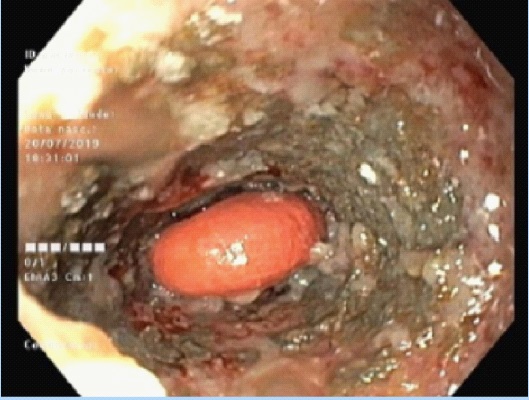
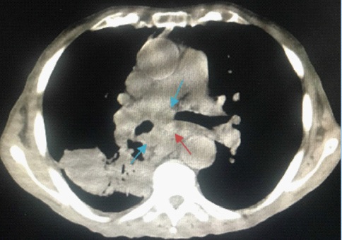
A 90-year old male diagnosed with stage III non-Hodgkin’s peripheral T-cell lymphoma under palliative treatment was admitted due to a feeling of food impaction and vomiting. He underwent upper digestive endoscopy which revealed an extensively necrotic esophageal mucosa with complete esophagus obstruction where two impacted pills were seen (Figure 1) and fragmented. Non-contrast chest computed tomography showed multiple exuberant mediastinal adenopathies conditioning extrinsic compression and esophageal collapse (Figure 2). Patient later underwent endoscopic reassessment which showed complete mucosa healing, therefore proceeding with esophageal stenosis endoluminal balloon dilation. Mediastinal adenopathies due to peripheral T cell-lymphoma are rare. This case represents an unusual example of extrinsic esophageal compression due to lymphoma leading to severe pill- induced esophagitis.