
Journal of Clinical Images and Medical Case Reports
ISSN 2766-7820
Clinical Image - Open Access, Volume 3
Radioopaque gall bladder stone detected on plain abdominal radiograph in an adult diabetic patient
Saurabh Puri, MBBS1*; Ashish Gupta, MBBS2
1Post Graduate Resident, Department of Internal Medicine, Max Super Specialty Hospital, Vaishali, UP, India.
2DNB general medicine, Senior Consultant, Department of Internal Medicine, Max Super Specialty Hospital, Vaishali, UP, India.
*Corresponding Author : Saurabh Puri
Post Graduate Resident, Department of Internal Medicine, Max Super Specialty Hospital, Vaishali, Ghaziabad, UP, India.
Email: Saurabhpuri119@gmail.com
Received : Aug 06, 2022
Accepted : Aug 31, 2022
Published : Sep 07, 2022
Archived : www.jcimcr.org
Copyright : © Puri S (2022).
Keywords: Gall bladder; Calculi; Radioopaque.
Citation: Puri S, Gupta A. Radioopaque gall bladder stone detected on plain abdominal radiograph in an adult diabetic patient. J Clin Images Med Case Rep. 2022; 3(9): 2036.
Description
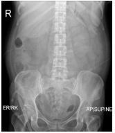
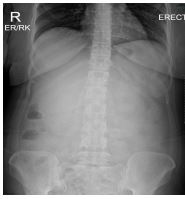
A 41-year-old female presented to emergency with complaints of recurrent vomiting, pain abdomen since last 24 hours. She is known case of diabetes, non-compliant to her medication. On examination, she was confused, GCS 11/15, and vitals were pulse rate 102/min, blood pressure 130/70 mmHg, respiratory rate 26/min, and random blood sugar 439 mg/dl. Arterial blood gas revealed pH 7.389, and urine routine revealed glucose 2 plus, ketone 1 plus. She was managed with intravenous fluids, insulin infusion, and other supportive measures. HbA1c was 12.4. X ray abdomen erect and supine were done which revealed two well defined oval radioopacities largest measuring 1.6 x 1.3 cm is noted in anticipated region of gall bladder fossa? calculi. USG whole abdomen confirmed cholelithiasis.
Discussion
Gallstones, also called cholelithiasis, are concretions that may occur anywhere within the biliary system, most commonly within the gallbladder, and is classified on the location of gallstones as cholecystolithiasis (gallstones within the gallbladder) and choledocholithiasis (gallstones within the bile ducts). Common risk factors include female sex, middle age, obesity, positive family history, recent rapid weight loss. Gallstones are symptomatic in 25% cases only with most common presentation being biliary colic. Gallstones are of three types i.e. cholesterol, pigmented and mixed [1].
Radiographically, ultrasound is gold standard for detecting gallstones [2]. Where as in 15-20% cases are of radiopaque gallstones seen in radiograph [3], and can be laminated/lamellated i.e. radiopaque outline with lucent center, as seen in our patient. Radiopaque Gall stone diagnosed through plain abdominal radiograph is unusual and rarely reported, making the image interesting and are of clinical importance, especially in diabetic patients.
References
- Chuang S, Hsi E, Lee K. Genetics of Gallstone Disease. Adv Clin Chem. 2013; 60: 143-185.
- Kothari S, Obinwanne K, Baker M, Mathiason M, Kallies K, et al. A Prospective, Blinded Comparison of Laparoscopic Ultrasound with Transabdominal Ultrasound for the Detection of Gallbladder Pathology in Morbidly Obese Patients. J Am Coll Surg. 2013; 216: 1057-1062.
- Bortoff G, Chen M, Ott D, Wolfman N, Routh W. Gallbladder Stones: Imaging and Intervention. Radiographics. 2000; 20: 751-766.