
Journal of Clinical Images and Medical Case Reports
ISSN 2766-7820
Clinical Image - Open Access, Volume 3
Dystrophic breast calcifications in a patient with
dermatomyositis
Yosra Bouattour1; Mouna Snoussi1*; Faten Frikha1; Raida Ben Salah1; Sameh Marzouk1; Zeineb Mnif2; Zouhir Bahloul1
1Internal Medicine Department, Hedi Chaker Hospital, Sfax Tunisia.
2Radiology Department, Hedi Chaker Hospital, Sfax Tunisia.
*Corresponding Author : Mouna Snoussi
Internal Medicine Department, Hedi Chaker Hospital, Sfax Tunisia.
Email: mounasnoussi23@gmail.com
Received : Sep 20, 2022
Accepted : Oct 20, 2022
Published : Oct 27, 2022
Archived : www.jcimcr.org
Copyright : © Snoussi M (2022).
Citation: Bouattour Y, Snoussi M, Frikha F, Salah RB, Marzouk S, et al. Dystrophic breast calcifications in a patient with dermatomyositis. J Clin Images Med Case Rep. 2022; 3(10): 2125.
Description
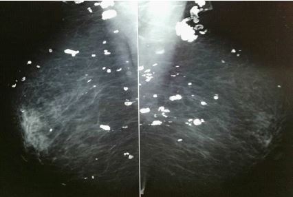
We report the case of a 34-year-old woman with a history of dermatomyositis in its typical form. She was treated by corticosteroids and methotrexate with no further relapses. She also had universalis calcinosis.
24 years later, the patient suffered from breast abscesses. Mammograms showed irregular, bizarre and coarse calcifications involving bilateral breasts. She received antibiotics for the infection.
Soft tissue calcifications in patients with dermatomyositis normally appear parallel to the long axes of muscles and interfascial planes [1]. Their localization in the breast’s subcutaneous tissue is uncommon described in few patients [2]. The diagnosis is based on mammography and ultrasonography.
References
- A Rhoden. Allowing repeated and Biomedical [1], we recognized. 765–773.
- V Singla, N Prabhakar, T Singh, A Sharma, N Khandelwal, et al. Mammography Findings of Breast Calcinosis in a Patient With Dermatomyositis. JCR J. Clin. Rheumatol. 2017; 23: 341.