Journal of Clinical Images and Medical Case Reports
ISSN 2766-7820
Case Report - Open Access, Volume 3
Active chorioretinal toxoplasmosis
Mehrdad Motamed Shariati; Sahel Khazaei*
Eye Research Center, Mashhad University of Medical Sciences, Mashhad, Iran.
*Corresponding Author : Sahel Khazaei, MD
Eye research center, Khatam Al-Anbia eye hospital, Mashhad University of Medical Sciences, Mashhad, Iran.
Tel: +98-918-645-4289;
Email: sahelkhazaei@yahoo.com
Received : Nov 10, 2022
Accepted : Dec 01, 2022
Published : Dec 08, 2022
Archived : www.jcimcr.org
Copyright : © Khazaei S (2022).
Citation: Shariati MM, Khazaei S. Active chorioretinal toxoplasmosis. J Clin Images Med Case Rep. 2022; 3(12): 2190.
Description
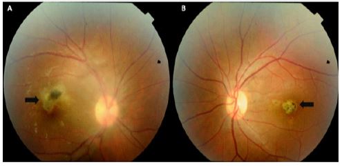
A systemically healthy 35-year-old female has complained of blurry vision of the Right Eye (RE) over the past week. The best-corrected visual acuity was 6/120 in both eyes. Mild conjunctival injection, fine keratic precipitates, and 1+ cell in the anterior chamber without hypopyon was detected in the RE. In dilated fundus examination of the RE, vitritis obscured portions of the retina and in the macular region, a creamy, grayish-white, raised retinal lesion was present adjacent to a chorioretinal scar (Figure 1A). In the left eye, two macular chorioretinal scars were seen (Figure 1B), findings that are consistent with chorioretinal toxoplasmosis. The diagnosis was supported by the results of the serologic evaluation. Toxoplasma gondii IgG and IgM antibodies were positive. Such lesions are typical of active chorioretinal toxoplasmosis. The patient was treated with intravitreal injection of clindamycin and dexamethasone, oral trimethoprim-sulfamethoxazole, and azithromycin. After 3 months complete healing of the active lesion occurred.