
Journal of Clinical Images and Medical Case Reports
ISSN 2766-7820
Clinical Image - Open Access, Volume 4
Takayasu’s arteritis diagnosed by carotid artery doppler
N Balamurugan, MD1; U Amaravathi, MD2; Swetha Ramesh, MBBS3
1Assistant Professor, Department of Emergency Medicine, JIPMER, Pondicherry, India.
2Senior Resident, Department of Emergency Medicine, JIPMER, Pondicherry, India.
3Junior Resident, Department of Emergency Medicine, JIPMER, Pondicherry, India.
*Corresponding Author : Swetha Ramesh
Junior Resident, Department of Emergency Medicine, JIPMER, Pondicherry- 605006, India.
Phone: +91-910-860-9203
Email: swetharamesh710@gmail.com
Received : Nov 10, 2022
Accepted : Dec 27, 2022
Published : Jan 03, 2023
Archived : www.jcimcr.org
Copyright : © Ramesh S (2023).
Citation:Balamurugan N, Amaravathi U, Swetha R. Takayasu’s arteritis diagnosed by carotid artery doppler. J Clin Images Med Case Rep. 2023; 4(1): 2226.
Description
TA is a large vessel vasculitis involving mainly the aorta and its primary branches resulting in segmental stenosis, occlusion or aneurysms [1]. Angiography is the gold standard for diagnosing TA but may miss wall changes in the early stages of disease [1]. So bedside ultrasonography can serve as a valuable tool in ED for diagnosing early TA. The role of B mode ultrasound for detection of arterial thickening and follow up of patients is supported by previous studies [2].
We report a case of a 27 years old female with no prior comorbidities who presented to the emergency department (ED) with complaints of gradual painless decline in her vision for three months. There was no history of trauma, fever, diplopia or headache. Her visual acuity was 20/100 in right eye and 20/200 in left eye. While assessing the vitals her upper limb pulses were not palpable. A thorough examination then revealed that she didn’t have bilateral carotid and upper limb pulses whereas all lower limb pulses were palpable. Ultrasound of her right common carotid artery showed Macaroni sign (Figure 1).
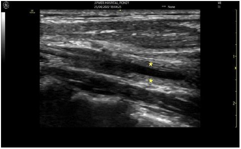

Ultrasound of her common carotid arteries showed smooth, homogenous, circumferential, echogenic wall thickening with stenosis (left > right). This finding, pathognomonic of Takayasu’s arteritis (TA), is the eponymous Macaroni Sign [3]. Fundus fluorescein angiography revealed neovascularization of disc with extensive microaneurysms in both eyes. CT angiography showed aortitis in the arch of aorta and its branches suggestive of TA (Figure 2). She underwent pan retinal photocoagulation and was started on steroids and is doing better.
References
- Ghembaza MEA, Boulenouar F, Lounici A. “Macaroni Sign” in Takayasu Arteritis. J Cardiovasc Imaging. 2018; 26: 186–7.
- Raninen RO, Kupari MM, Pamilo MS, Pajari RI, Poutanen VP, Hekali PE. Arterial wall thickness measurements by B mode ultrasonography in patients with Takayasu’s arteritis. Annals of the rheumatic diseases. 1996; 55(7): 461-5.
- Maeda H, Handa N, Matsumoto M, Hougaku H, Ogawa S, Oku N, et al. Carotid lesions detected by B-mode ultrasonography in Takayasu’s arteritis: “macaroni sign” as an indicator of the disease. Ultrasound Med Biol. 1991; 17:695–701.